Figure 5. The effect of CX46A397fs mutation on the secondary structure of GJA3 protein. A: The frameshift prediction results using the GOR method. The changes in secondary structure specific in CX46S380fs are indicated
in red (along with a specific change in the secondary structure of one out of the 17 amino acids); the changes in secondary
structure shared by CX46A397fs and CX46S380fs are indicated in blue. One amino acid at the 398th site of CX46A397fs and CX46S380fs mutant possesses a secondary structure different from that of the reference, indicated
in brown and blue, respectively. B: The alignment of CX46A397fs and CX46S380fs mutant protein sequences demonstrated 96.8% identity in 466 amino acid residues.
The 17 amino acids that differ between CX46S380fs and CX46A397fs are blanketed in red. The 29 amino acids that are shown to
impair the function of CX46380fs mutant are blanketed in black.
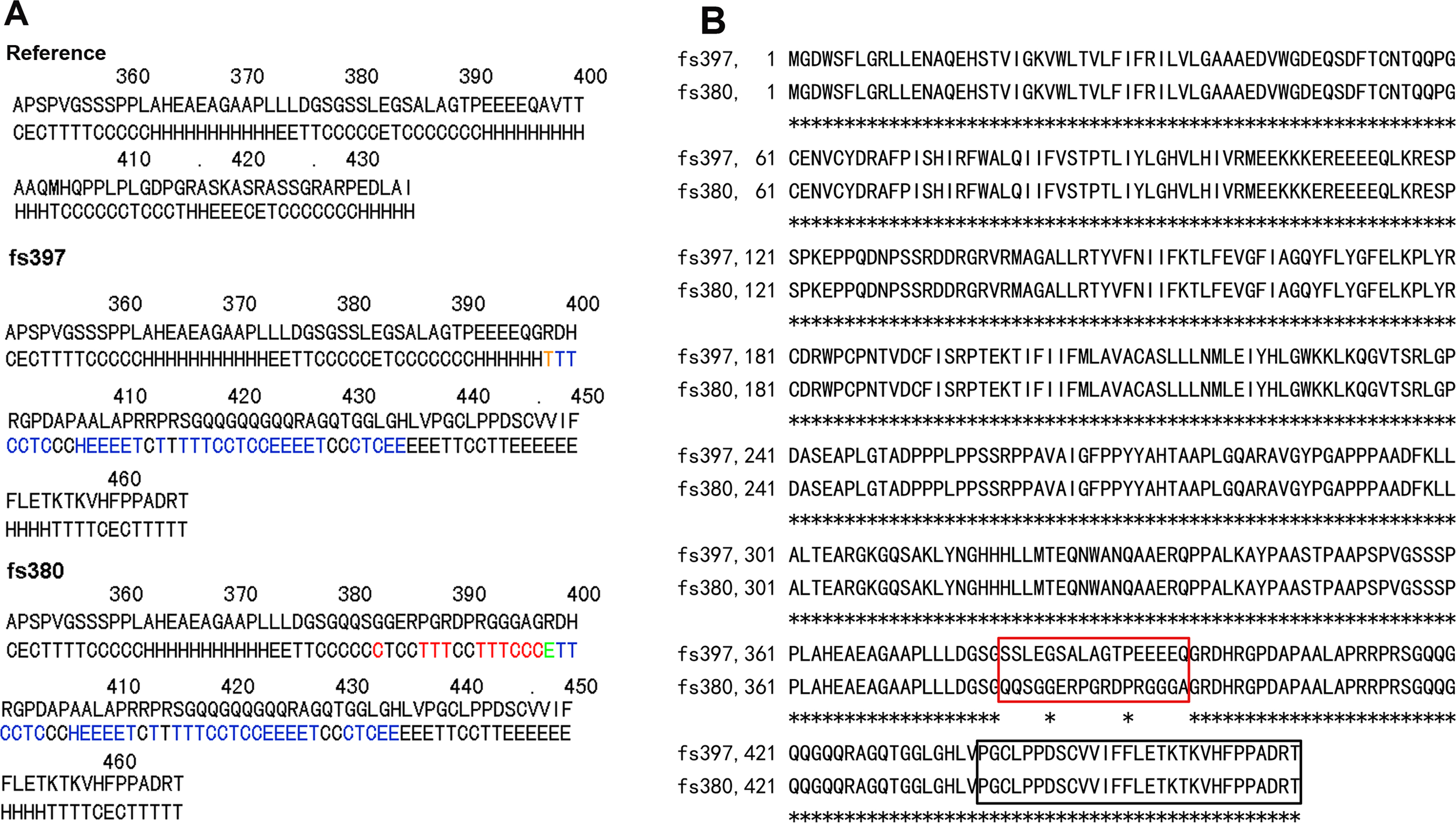
 Figure 5 of
Zhou, Mol Vis 2013; 19:789-795.
Figure 5 of
Zhou, Mol Vis 2013; 19:789-795.