Figure 3. Neovascularization under relative hypoxic conditions in C57BL/6J mouse retinas. After the mice were relocated to room air
for 24 h, the size of the vasoobliteration (VO) area decreased slightly, but the deep vascular plexus was observed in the
peripheral area, in which area superficial vessels were preserved during hyperoxia. At P15, the size of the VO area continued
to decrease, and the deep—but not the intermediate—vascular plexus was observed in the area where superficial vessels were
preserved during hyperoxia. The retinal arteries and veins had a dilated and tortuous appearance, and vascular branch sprouts
emerged from existing vessels. From P17 to P19, retinal neovascularization progressed rapidly, and numerous vascular sprouts
appeared in the transition zone between the vascular and avascular retina. The deep and intermediate plexuses can be observed
in almost the same area as the superficial plexus. From P21 to P23, the size of the VO area was small, and neovascularization
began to regress. At P25, the VO area was fully revascularized in three layers, neovascularization had completely resolved,
and the retinal vessels began to remodel.
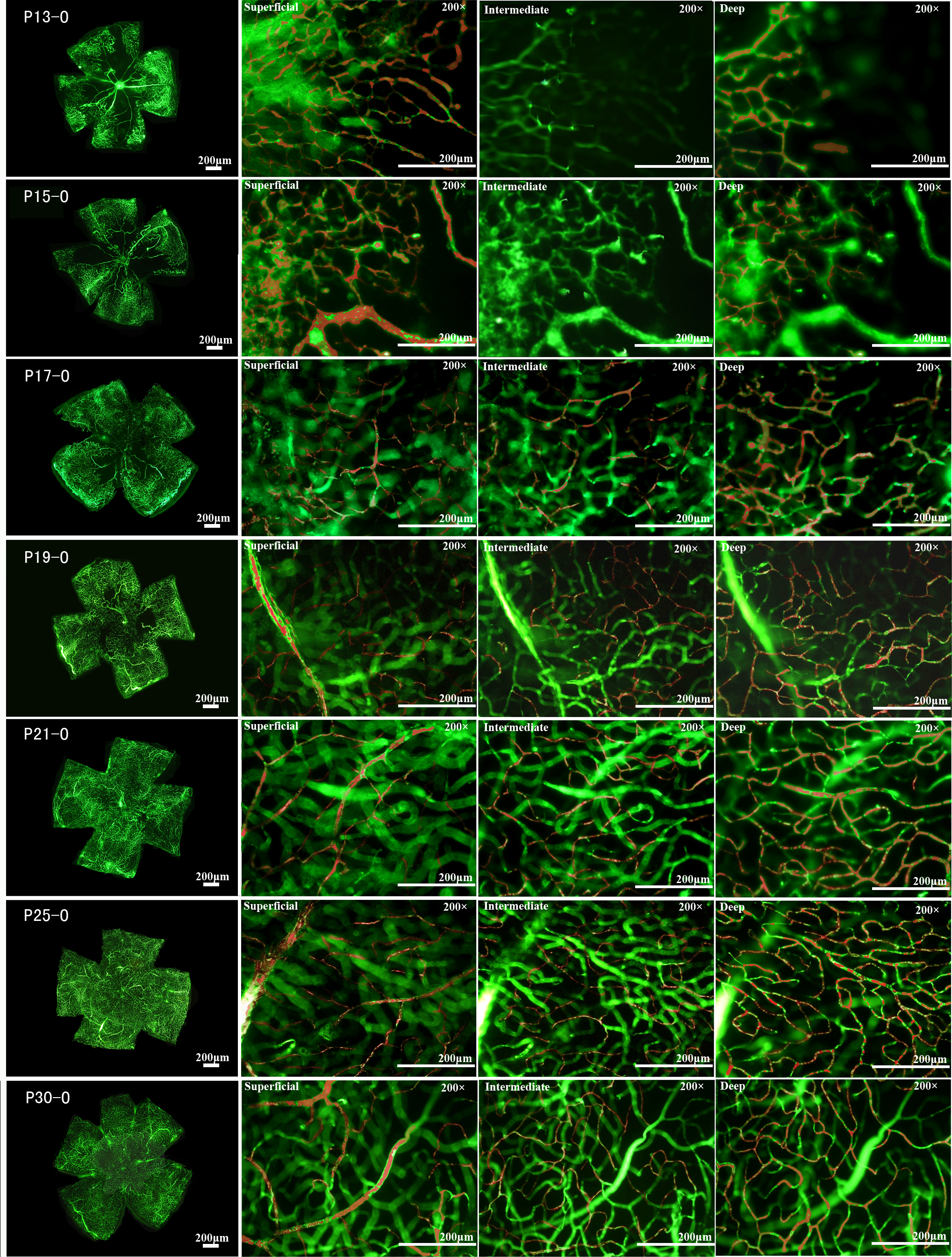
 Figure 3 of
Yang, Mol Vis 2013; 19:775-788.
Figure 3 of
Yang, Mol Vis 2013; 19:775-788.