Figure 10. Electrophoretic mobility shift assay of STAT1 activation. A: A putative STAT-binding element (shown in dark background) that is evolutionarily conserved in mammalian species was detected
in the promoter region of MIR146B. The dashed line indicates the sequence of the oligonucleotide probe used for the electrophoretic mobility shift assay. B: Electrophoretic mobility shift assay shows that the putative MIR146B STAT-binding element lacks the ability to bind STAT1. The assay was performed with biotin-labeled oligonucleotides containing
either a known STAT1-binding site (shown as STAT1, left side of the blot in the figure) or the putative STAT-binding element
of MIR146B (shown as MIR146B, right side of the blot in the figure) and using nuclear extracts from human retinal pigment epithelial
(HRPE) cells. The arrow identifies the STAT1 protein binding. Lane P, probe alone; lane C, extract from control cells; lane
I, extract from cells treated with interferon (IFN)-γ; lane M, extract from cells treated with a mixture of IFN-γ, tumor necrosis
factor (TNF)-α, and interleukin (IL)-1β; lane U, a 50-fold excess of corresponding unlabeled oligonucleotide and extract from
cells treated with a mixture of IFN-γ, TNF-α, and IL-1β. The data from one of the two similar experiments are shown.
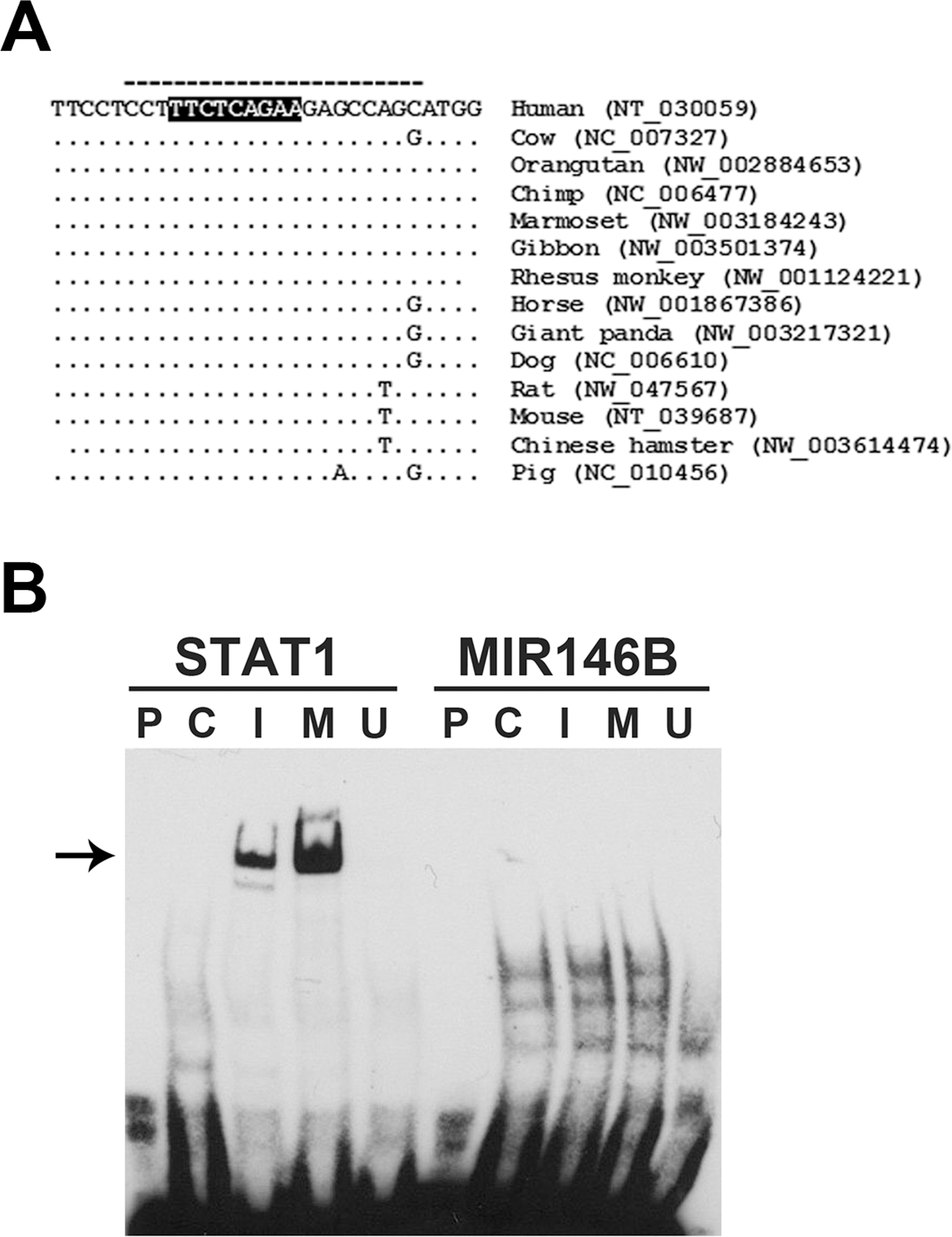
 Figure 10 of
Kutty, Mol Vis 2013; 19:737-750.
Figure 10 of
Kutty, Mol Vis 2013; 19:737-750.