Figure 1. Human retinal pigment epithelial cells responded to proinflammatory cytokines by markedly altering the expression of several
genes. The cells were treated with a cytokine mixture consisting of interferon (IFN)-γ (100 u/ml), tumor necrosis factor (TNF)-α
(10 ng/ml), and interleukin (IL)-1β (10 ng/ml) for 16 h. Total RNA fractions isolated from control and treated cells were
reverse-transcribed and the gene expression analyzed by real-time PCR using the relative quantification method. The mean CT values were as follows: control=26.95±0.02 and treated=19.84±0.04 for CCL2; control=33.04±0.08 and treated=17.96±0.02 for
CCL5; control=32.89±0.09 and treated=18.41±0.04 for CXCL9; control=31.95±0.02 and treated=17.23±0.02 for CXCL10; control=30.93±0.02
and treated=18.79±0.01 for IL-6; control=24.95±0.02 and treated=27.45±0.02 for HMOX1; and control=19.53±0.07 and treated=18.94±0.03
for GAPDH (endogenous control). Expression of CCL2, CCL5, CXCL9, CXCL10, and IL-6 was increased and that of HMOX1 decreased
following the treatment; p<0.001, n=3. The data shown are representative of three different experiments.
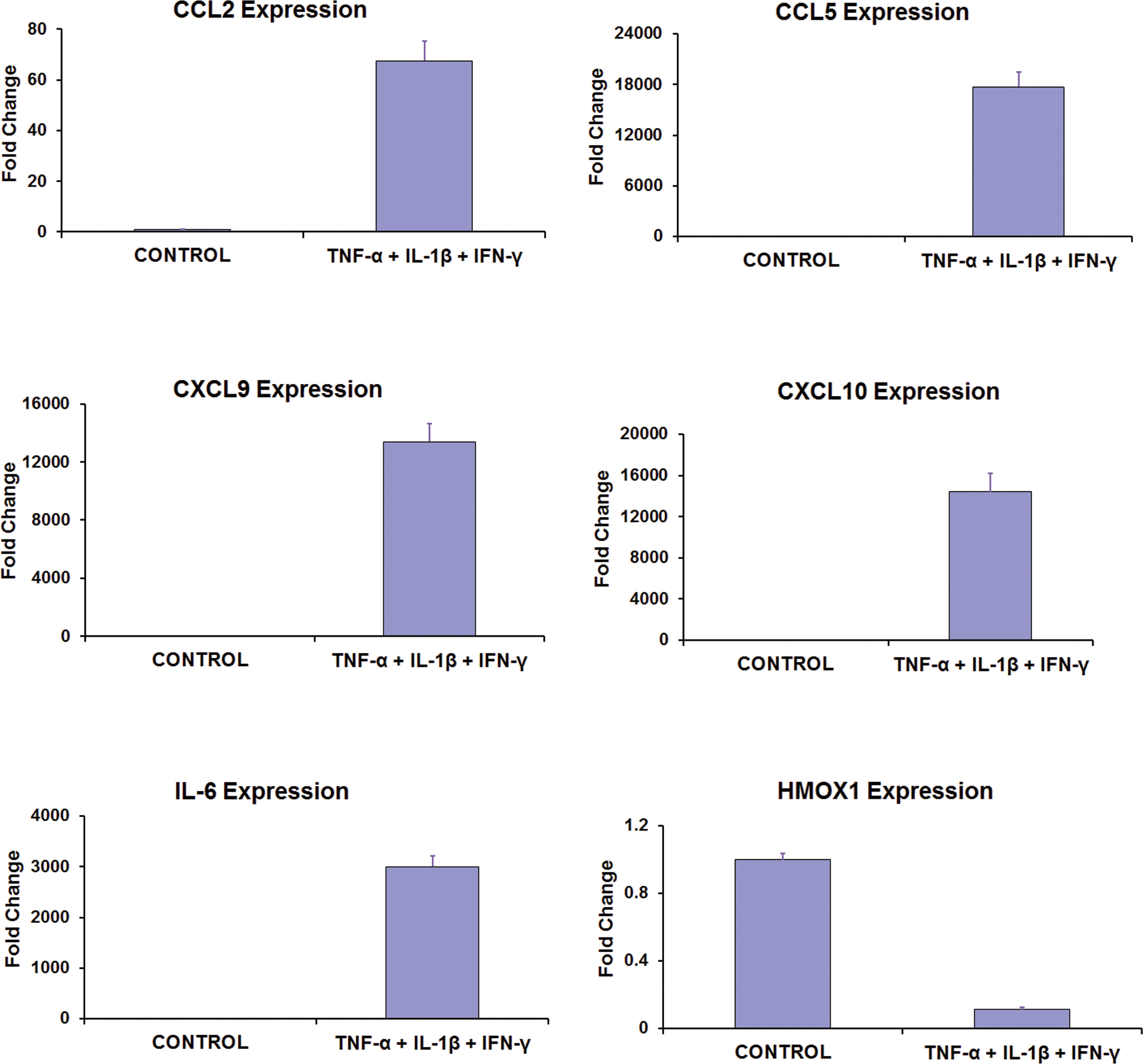
 Figure 1 of
Kutty, Mol Vis 2013; 19:737-750.
Figure 1 of
Kutty, Mol Vis 2013; 19:737-750.