Figure 2 of
Liu, Mol Vis 2013; 19:695-701.
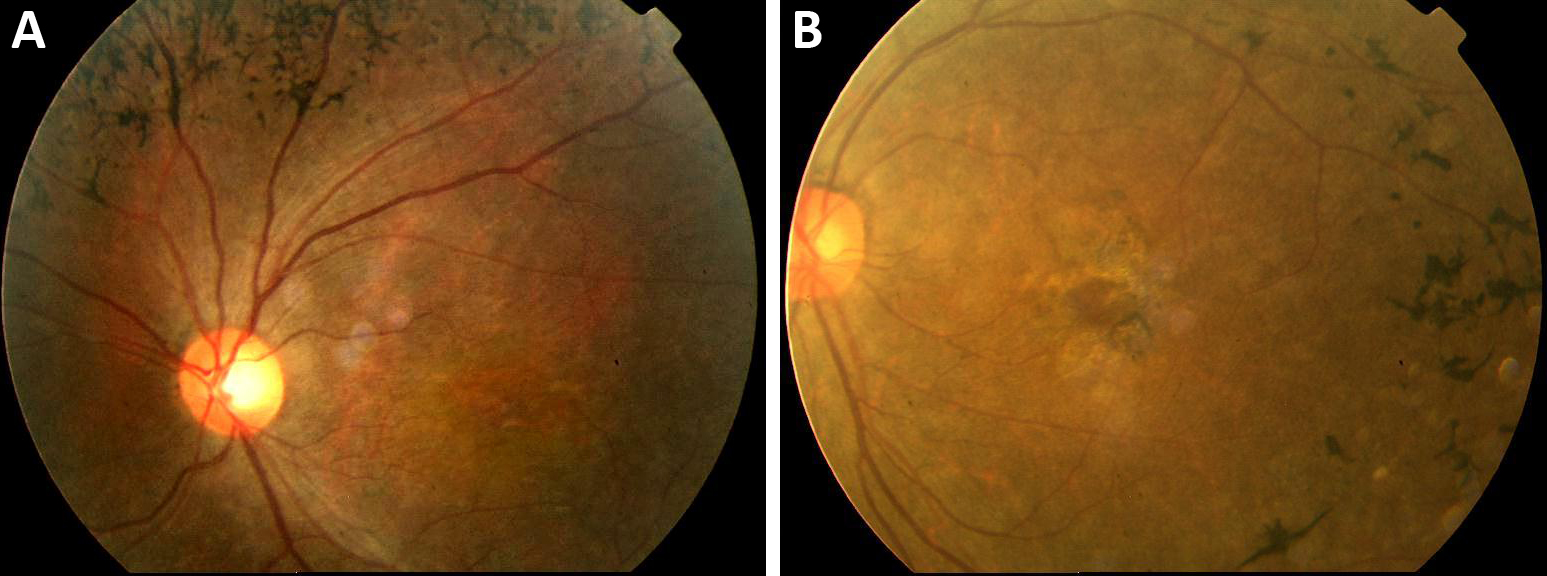
Figure 2.
Fundus photographs of the proband at age 20 years.
A
: Retinal vessel attenuation and characteristic bone spicule pigment are shown, indicating a typical retinitis pigmentosa phenotype.
B
: The macula was involved.
 Figure 2 of
Liu, Mol Vis 2013; 19:695-701.
Figure 2 of
Liu, Mol Vis 2013; 19:695-701.