Figure 3. Immunocytochemistry of limbal epithelial cells cultured in supplemental hormonal epithelial medium (SHEM), keratinocyte serum-free
medium (KSFM) and Epilife showing the number of positive cells (N cell +) and percentage of positive cells. The Kruskal–Wallis
test was performed. There were significant differences for p63. (Light microscope 40x).
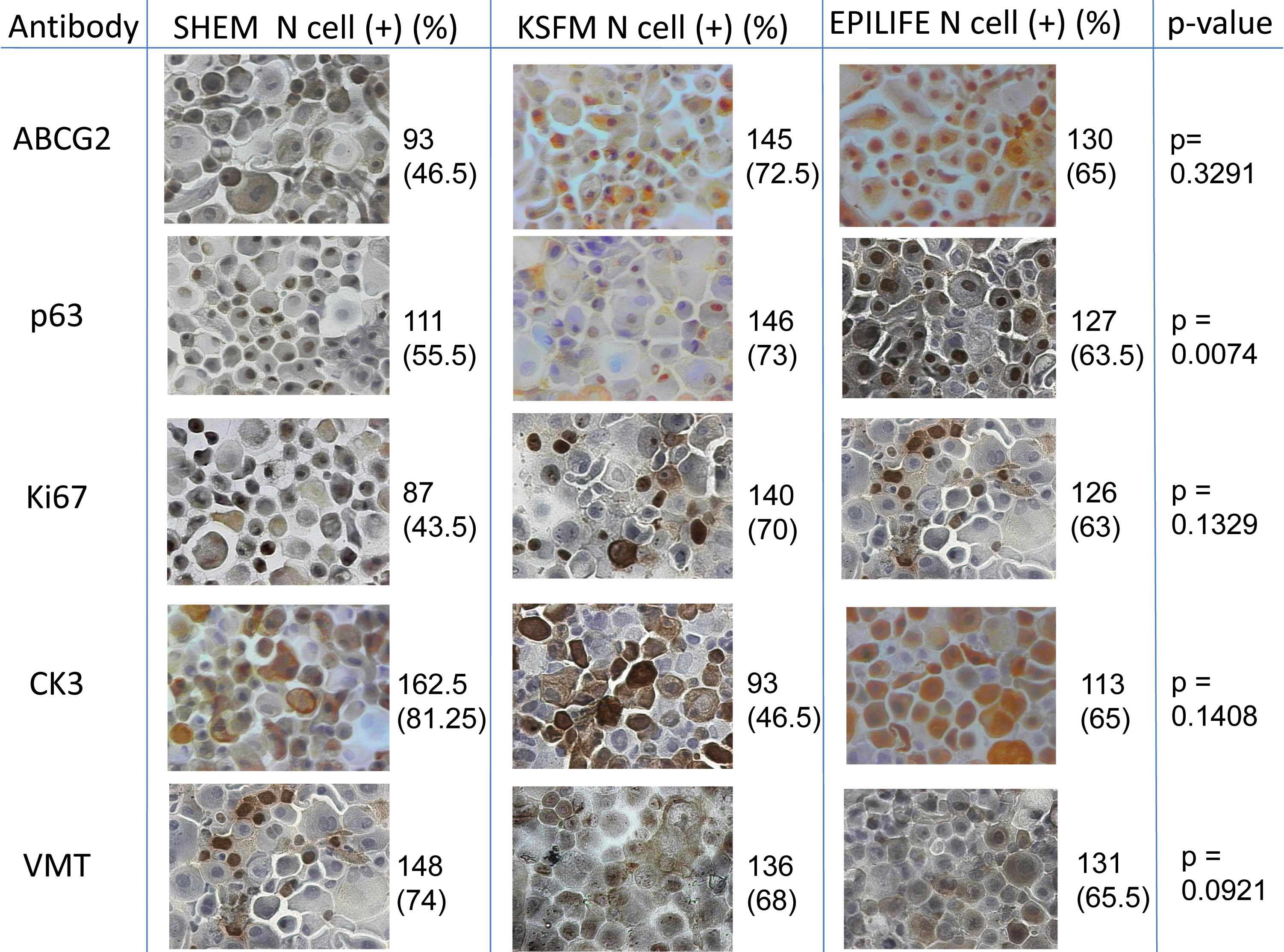
 Figure 3 of
Loureiro, Mol Vis 2013; 19:69-77.
Figure 3 of
Loureiro, Mol Vis 2013; 19:69-77.