Figure 2. Plot of cell growth area over time for limbal epithelia cultivated in the culture media supplemental hormonal epithelial medium
(SHEM), keratinocyte serum-free medium (KSFM), and Epilife. There were significant differences in cell growth area in SHEM
and KSFM media at 10, 12, and 15 days of culture. One-way ANOVA was performed. Bars represent the standard deviation.
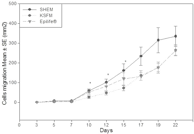
 Figure 2 of
Loureiro, Mol Vis 2013; 19:69-77.
Figure 2 of
Loureiro, Mol Vis 2013; 19:69-77.