Figure 1 of
Loureiro, Mol Vis 2013; 19:69-77.
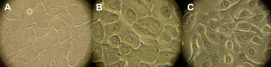
Figure 1.
Optical morphological analysis of the limbal epithelial cells cultivated in three different culture media.
A
: supplemental hormonal epithelial medium (SHEM),
B
: keratinocyte serum-free medium (KSFM), and
C
: Epilife. Light microscope=40×.
 Figure 1 of
Loureiro, Mol Vis 2013; 19:69-77.
Figure 1 of
Loureiro, Mol Vis 2013; 19:69-77.