Figure 6. Immunolocalization of collagen IV (ColIV) in the iris of 1- to 2-month-old mice. Positive expression can be seen in the lens
capsule (lc) in all groups of mice. Expression in the iris (i) and Descemet’s membrane (dm) is organized in the wild-type
(WT) and matrix metalloproteinase-9 knockout (MMP-9KO) mice. Expression is also apparent in the transgenic (TG) and TG/MMP-9KO
mice. However, it is distorted, consistent with the tissue distortion in these animals. 20X, bar=100 µm.
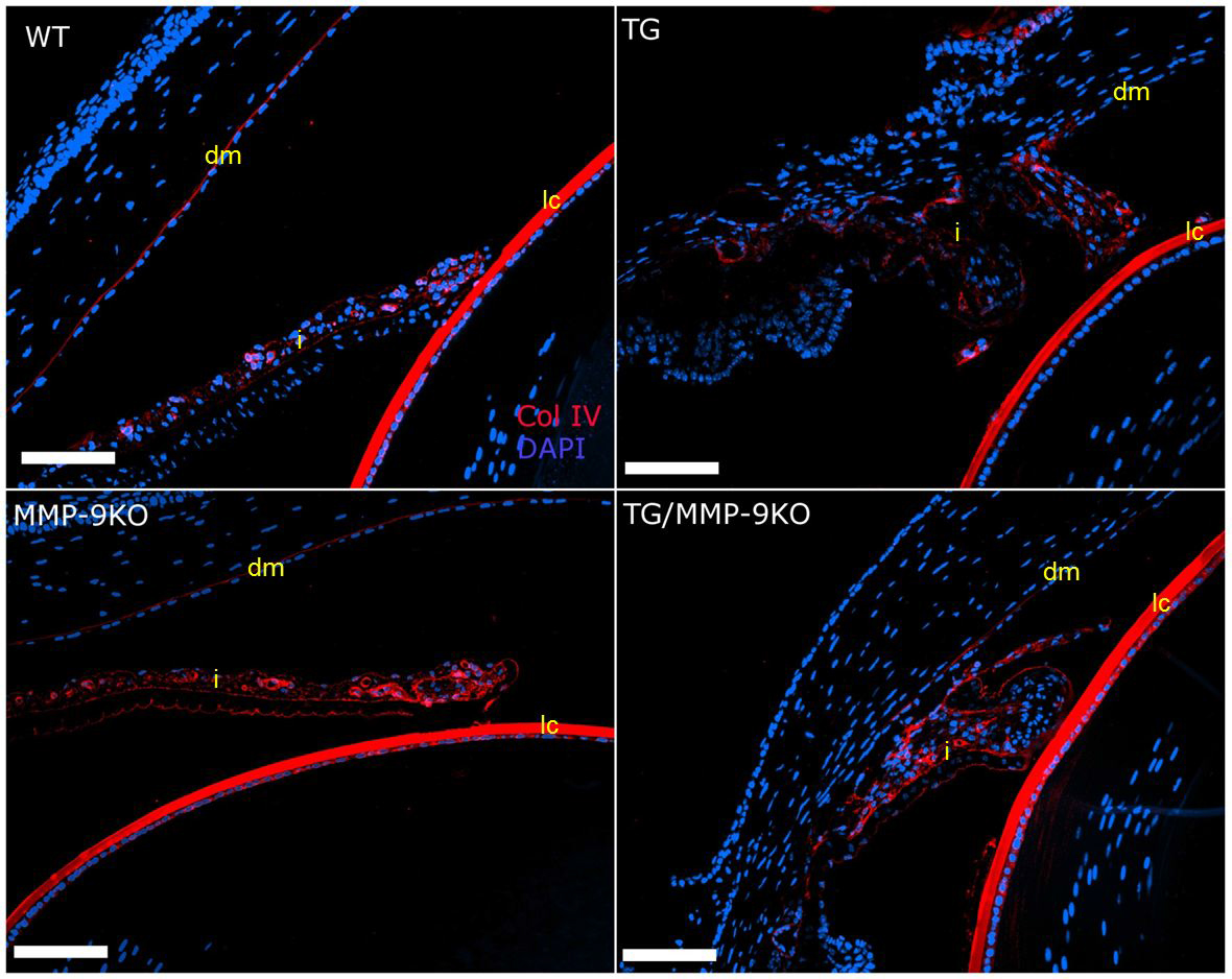
 Figure 6 of
Robertson, Mol Vis 2013; 19:684-695.
Figure 6 of
Robertson, Mol Vis 2013; 19:684-695.