Figure 5. Immunolocalization of collagen IV (ColIV) in the ciliary body and angle of 1- to 2-month-old mice. Organized expression of
ColIV can be seen in the trabecular meshwork (tm; region designated by the yellow arrowheads) and throughout the rays of the
ciliary body (cb) in the open angles (yellow dashed line) of the wild-type (WT) and matrix metalloproteinase-9 knockout (MMP-9KO)
mice. In the transgenic (TG) and TG/MMP-9KO mice, ColIV expression appears less organized. 40X, bar=50 µm.
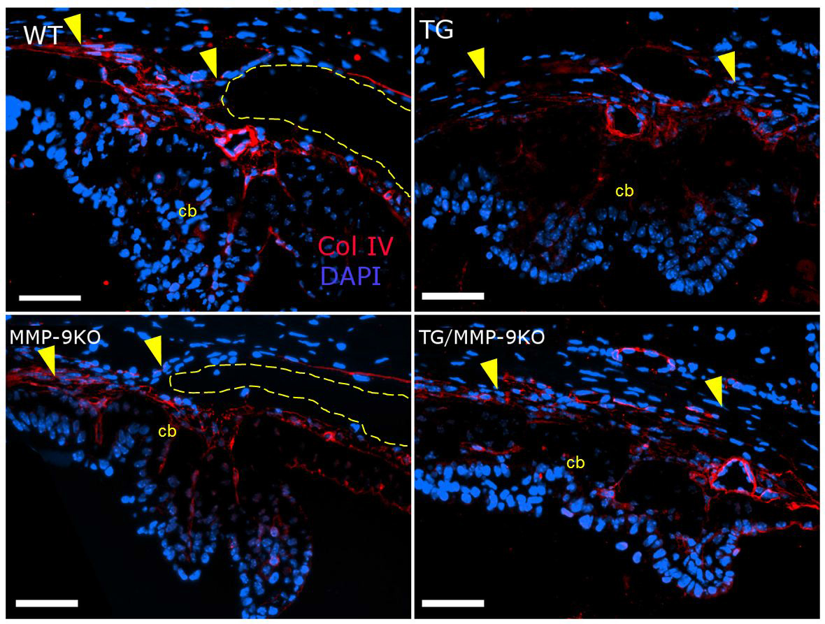
 Figure 5 of
Robertson, Mol Vis 2013; 19:684-695.
Figure 5 of
Robertson, Mol Vis 2013; 19:684-695.