Figure 2. Hematoxylin and eosin–stained sections from 1- to 2-month-old mice. Adhesions between the iris (i) and corneal endothelium
(ce) are easily visible in the transforming growth factor beta (TGFβ) transgenic (TG) and matrix metalloproteinase-9 knockout
(MMP-9KO)/TGFβ1 transgenic mice but are absent in the wild-type (WT) and MMP-9KO mice. 5X, bar=200 µm.
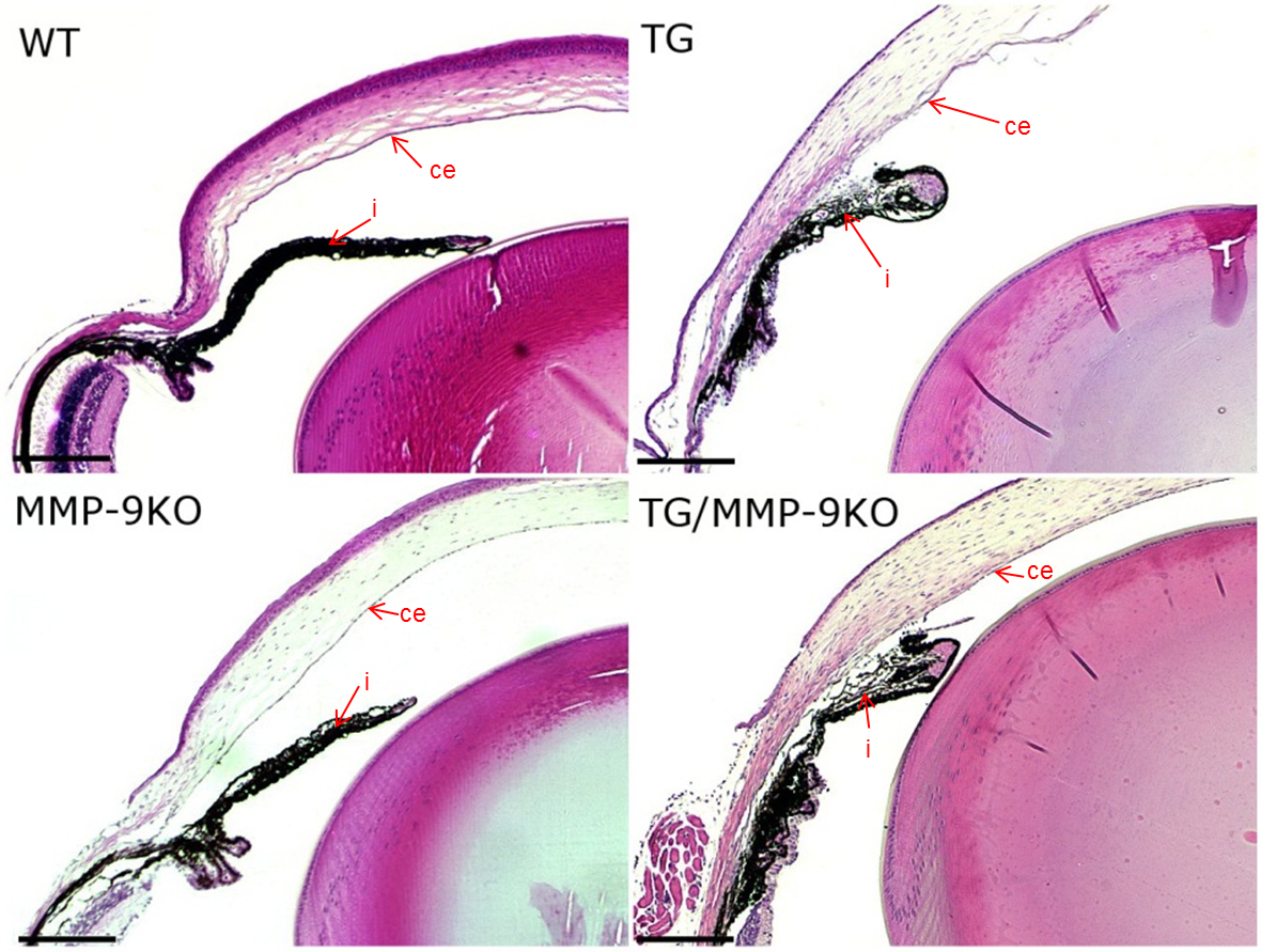
 Figure 2 of
Robertson, Mol Vis 2013; 19:684-695.
Figure 2 of
Robertson, Mol Vis 2013; 19:684-695.