Figure 2. Fundus photographs of affected and healthy individuals. A, B: Fundus photographs of family A, proband IV:5, show bone spicules, retinal vessel attenuation, and macular degeneration.
C, D: Fundus photographs of the proband’s sister (IV:4) show the salt-and-pepper appearance of both fundi, the presence of bone
spicules in the midperiphery, and bull’s eye macular atrophy (indicated by the arrows). E, F: Fundus photograph of family B proband IV:5 reveals pigmentary deposits and retinal vessel attenuation. G, H: Fundus photographs of a healthy individual from family A (IV:6).
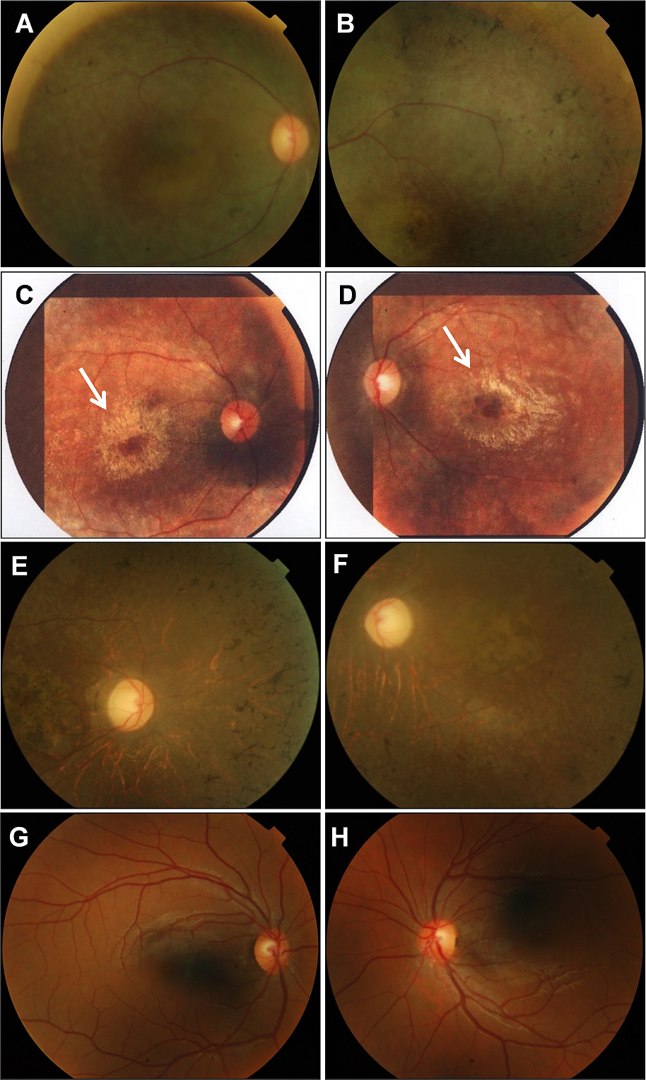
 Figure 2 of
Ajmal, Mol Vis 2013; 19:644-653.
Figure 2 of
Ajmal, Mol Vis 2013; 19:644-653.