Figure 2. Comparison of porcine corneal epithelial cells (A–C) with primary human corneal epithelial cells (D–F), magnification 250X, and of primary porcine corneal keratocytes (G–I) with primary human corneal keratocytes (J–L), magnification 50X. Alpha 2,3 sialic acid terminal saccharides were stained with fluorescein isothiocyanate–labeled Maackia amurensis I lectin (green, A, D, G, J), and the nuclei were counterstained with 4', 6-diamidino-2-phenylindole (blue, B, E, H, K). C, F, I, and L show the merged images.
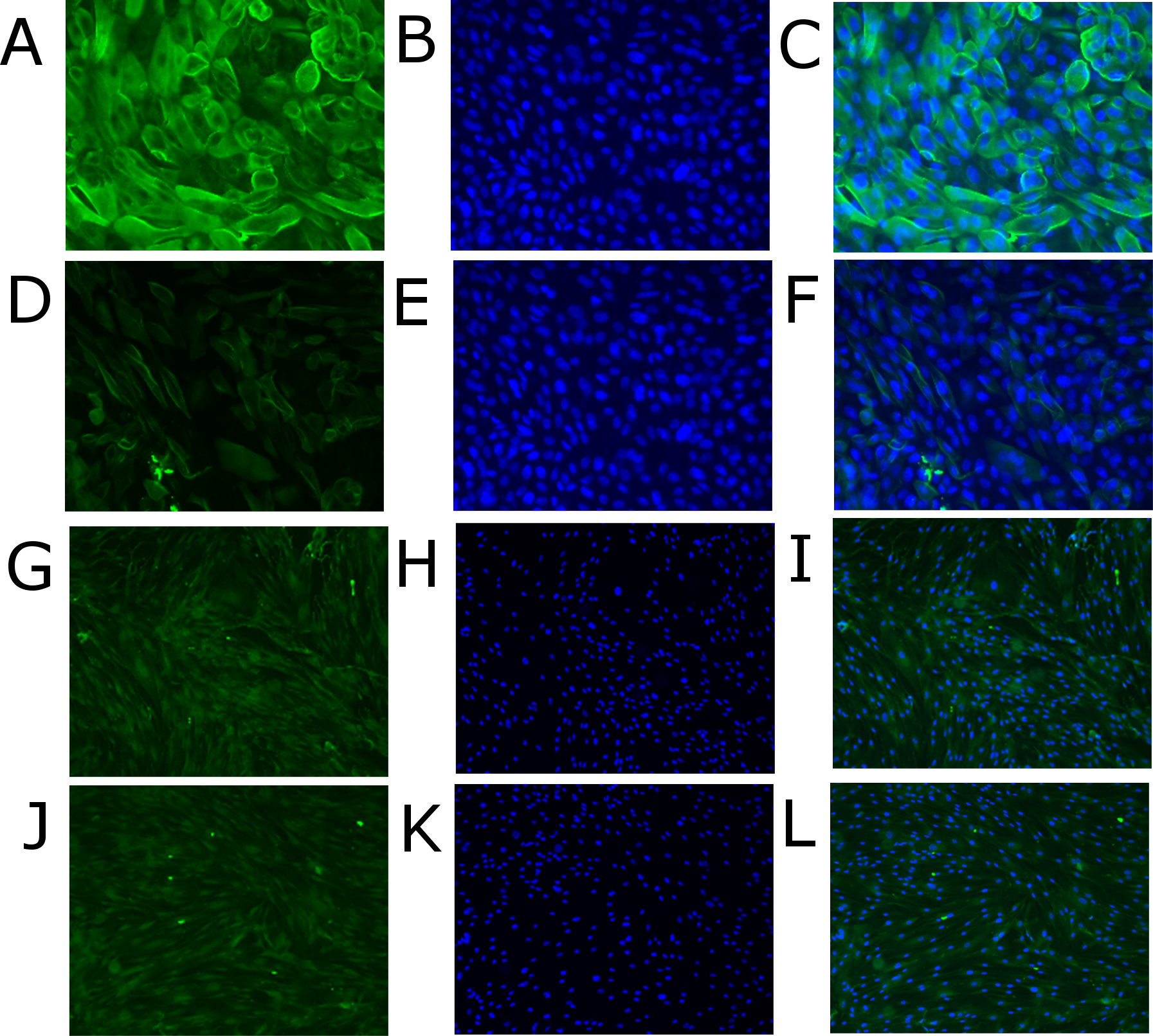
 Figure 2 of
Ramke, Mol Vis 2013; 19:614-622.
Figure 2 of
Ramke, Mol Vis 2013; 19:614-622.