Figure 1 of
Ramke, Mol Vis 2013; 19:614-622.
Figure 1.
Phase contrast microscopy of primary porcine corneal epithelial cells (PPCE;
A
), primary human corneal epithelial cells (PHCE;
B
), primary porcine corneal keratocytes (PPCK;
C
), and primary human corneal keratocytes (PHCK;
D
).
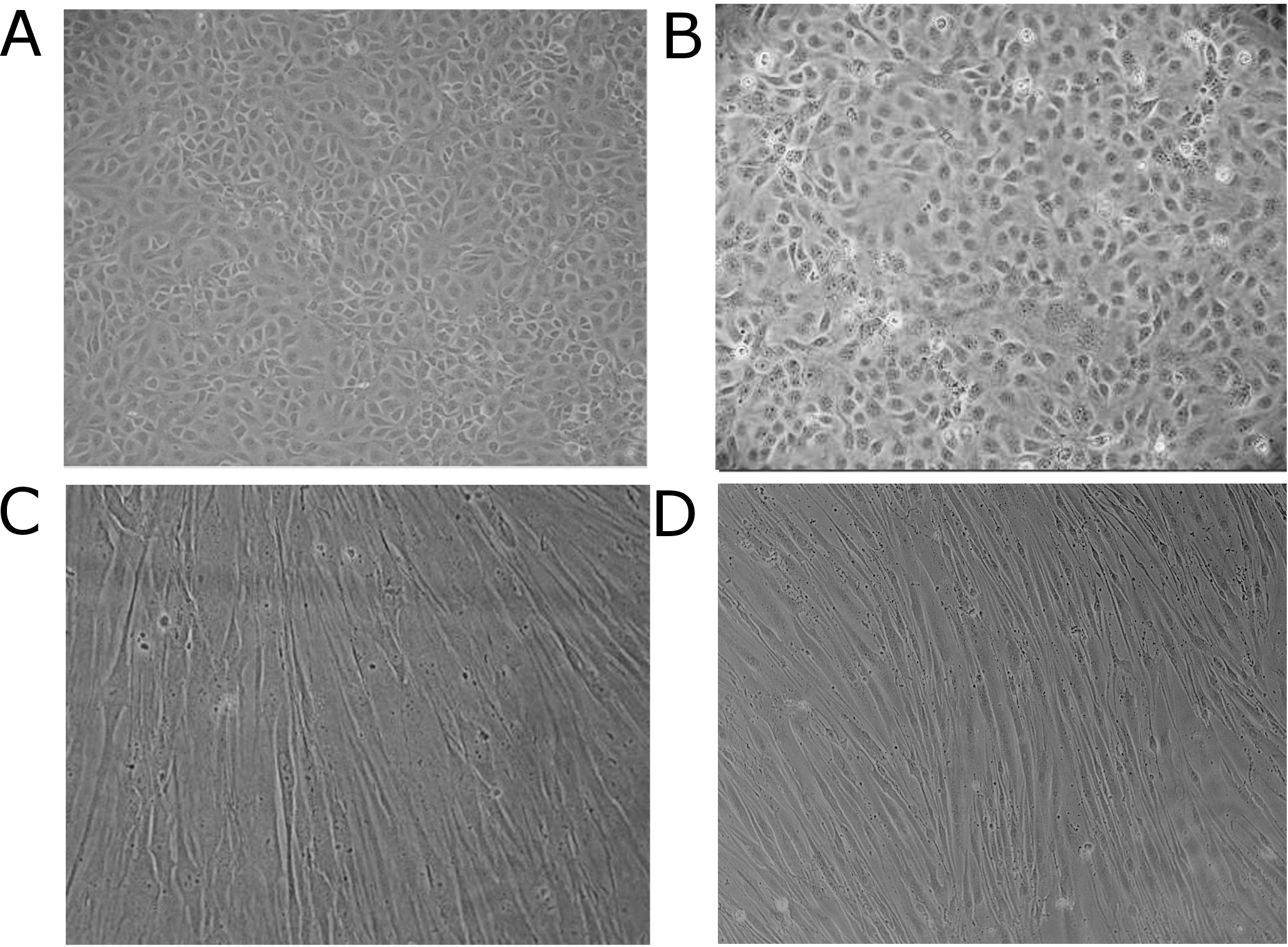
 Figure 1 of
Ramke, Mol Vis 2013; 19:614-622.
Figure 1 of
Ramke, Mol Vis 2013; 19:614-622.