Figure 3. Keratin 17 (K17) expression was increased in the epithelium of the pterygium. A-F demonstrates representative images of immunohistochemical staining of K17 in the normal conjunctiva (A, B), primary pterygium (C, D), and recurrent pterygium (E,F); A, C, E demonstrate representative images of H&E staining indicating the nuclei in the normal conjunctiva and the pterygium. Expression
of K17 in the basal layer of epithelium cells was increased (D, F; as arrows indicated), compared to the normal conjunctiva (B). G and I demonstrate representative images and statistical analysis of western blotting of K17 in the normal conjunctiva, primary
pterygium, and recurrent pterygium. The K17 protein level was statistically significantly increased in the primary pterygium
and the recurrent pterygium, compared to the normal conjunctiva. Data are represented as mean±SEM, n=3, **: p<0.01; ***:p<0.001
versus conjunctiva. H demonstrates quantitative real-time PCR data of K17mRNA in normal conjunctiva, primary pterygium, and recurrent pterygium.
The K17 mRNA level was statistically significantly upregulated in the primary and recurrent pterygium. Data are represented
as mean±SEM, n=10 in each group, *** p<0.001 versus recurrent pterygium. Scale bar: 100 μm.
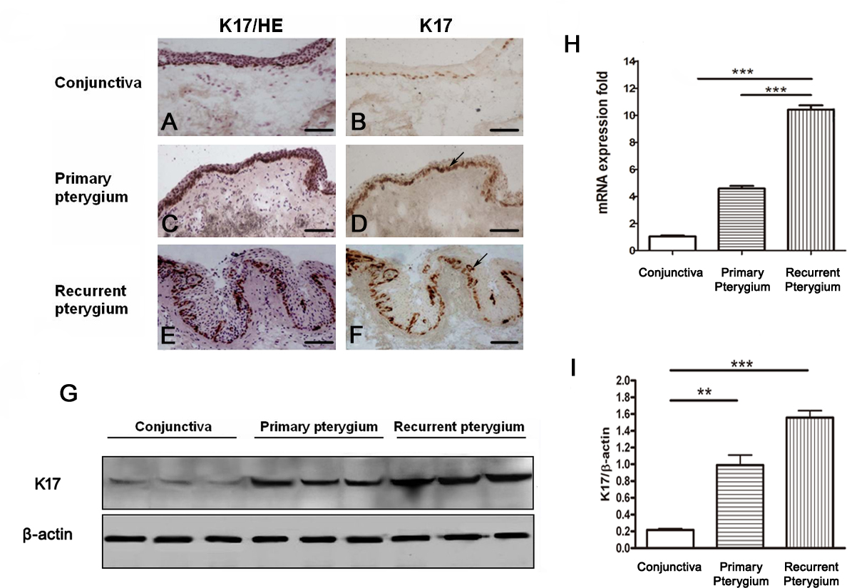
 Figure 3 of
Xu, Mol Vis 2013; 19:604-613.
Figure 3 of
Xu, Mol Vis 2013; 19:604-613.