Figure 3. Representative light microscopy pictures of staining of B lymphoma Mo-MLV insertion region 1 (BMI-1) antigen in human retinoblastoma
retinae. (Upper panel) Undifferentiated retinoblastomas and (lower panel) differentiated retinoblastomas grouped according
to TNM classification. Some specimens had an indistinct inner nuclear layer (marked by red brackets). There was no consistent
difference of BMI-1 expression in retinas of undifferentiated and differentiated retinoblastomas. Scale bars: 50 μm.
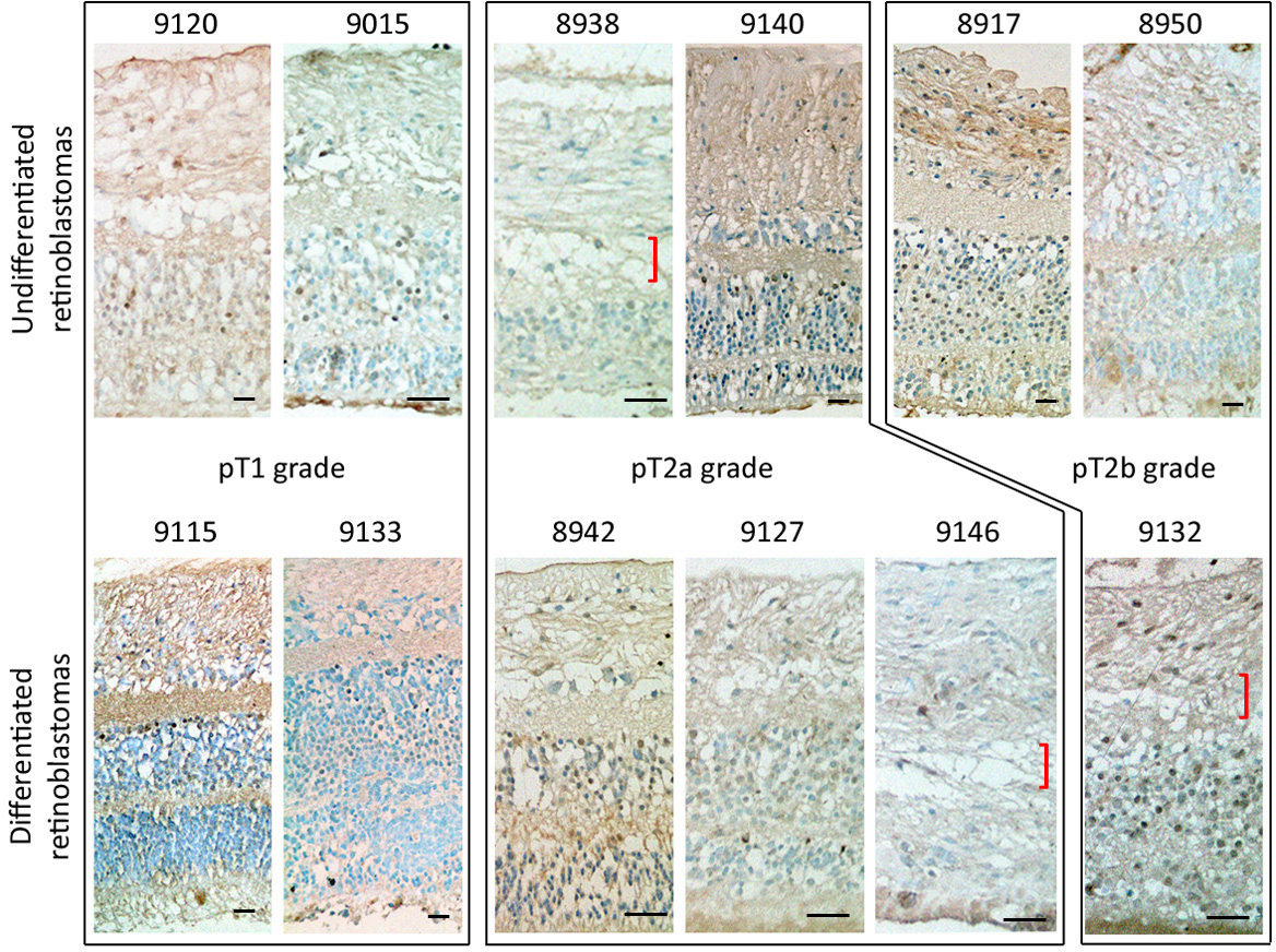
 Figure 3 of
Ren, Mol Vis 2013; 19:561-574.
Figure 3 of
Ren, Mol Vis 2013; 19:561-574.