Figure 2. Representative light microscopy pictures of staining of B lymphoma Mo-MLV insertion region 1 (BMI-1) antigen in differentiated
retinoblastomas grouped according to Tumor, Nodes, Metastasis (TNM) classification. A: Hematoxylin and eosin staining. Closed red arrows in A show the Flexner-Wintersteiner rosettes. B: Magnified images in C: weak to moderate BMI-1 expression in differentiated retinoblastomas. D: Background staining of BMI-1 in retinoblastoma 8927 with incubation of primary antibody. E: Negligible BMI-1 expression in a normal human retina section. Scale bars: 50 µm.
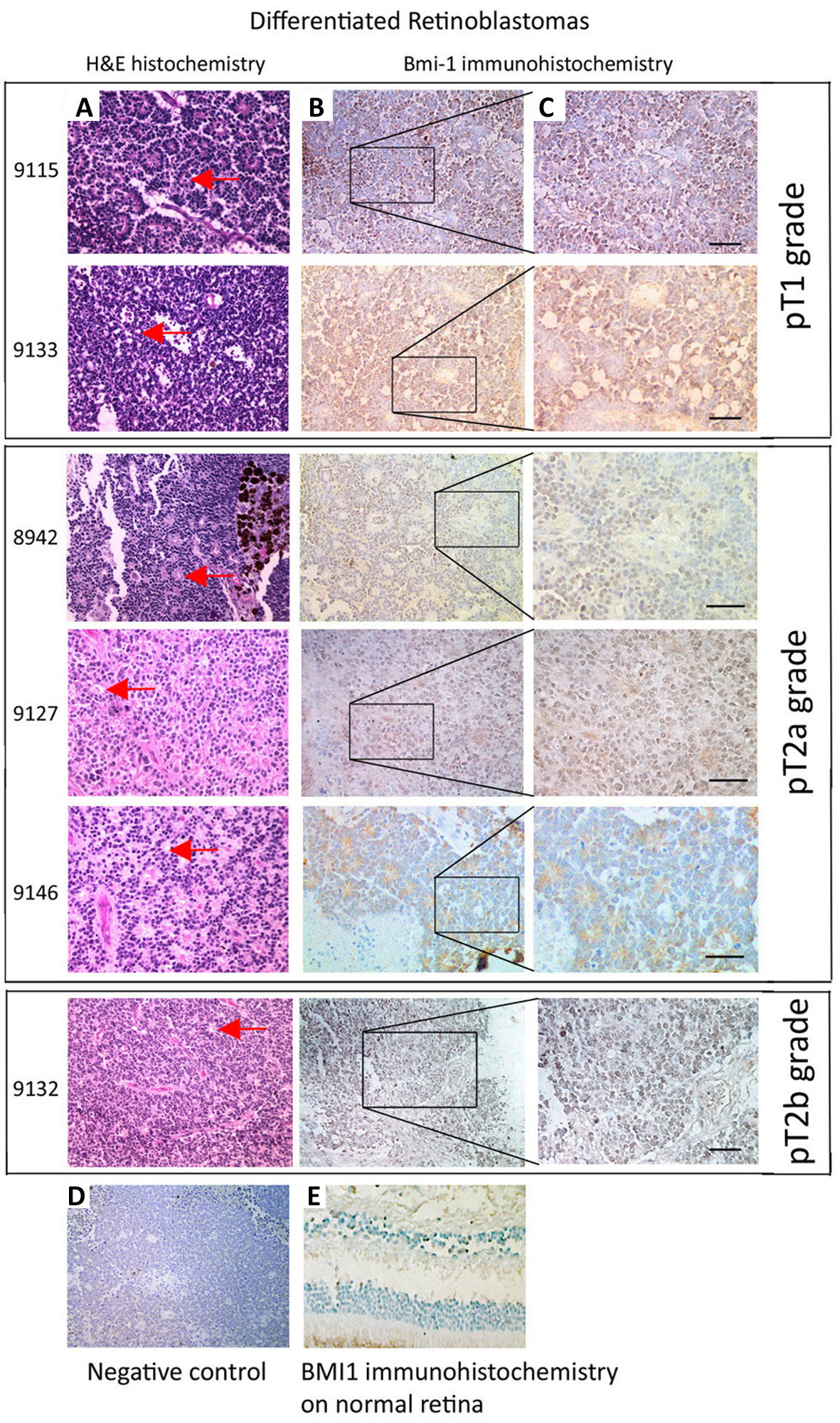
 Figure 2 of
Ren, Mol Vis 2013; 19:561-574.
Figure 2 of
Ren, Mol Vis 2013; 19:561-574.