Figure 5. Retinal damage at the laser site at d12. Two days after photodynamic therapy (PDT). The retina was perfused with FITC-dextran
and stained with TRITC-lectin. The site of the choroidal neovascularization (CNV) laser treatment in the center of the picture
(dashed circle) is not perfused after treatment with Visudyne and PDT though modified capillaries are there as detected with
lectin staining. This effect was not found when Visudyne was substituted by cationic liposomes (CL)-VTP or in the controls.
Dextran was washed out during lectin staining from the large vessels only but remained in the capillaries. The focus is on
the deep retinal vascular network, and the blurred green capillaries are connections to the superficial vascular net that
are out of focus.
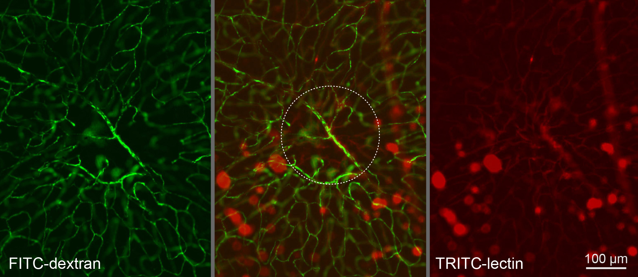
 Figure 5 of
Gross, Mol Vis 2013; 19:54-61.
Figure 5 of
Gross, Mol Vis 2013; 19:54-61.