Figure 3. Scanning laser microscopy of choroidal neovascularization (CNV). A laser site 14 days after laser coagulation shows a central
hole in the choroid and retinal pigment epithelium (RPE) from which a bundle of vessels is penetrating the retina (retina
was removed during preparation).
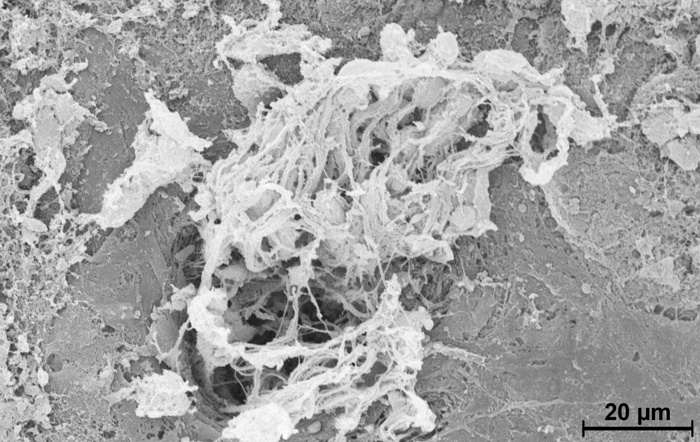
 Figure 3 of
Gross, Mol Vis 2013; 19:54-61.
Figure 3 of
Gross, Mol Vis 2013; 19:54-61.