Figure 2. Effect of cationic liposomes on choroidal neovascularization (CNV). CNV was induced by laser coagulation. Test and control
substances were injected in the tail vein every second day. Ten days after laser coagulation, mice were perfused with FITC-dextran
and scleral flatmounts were prepared. A: While trehalose resulted in a large circular neovascularized area (surrounded by a red dashed line), the CNV of cationic
liposomes (CL) and Taxol was smaller. EndoTAG-1 and LipoSPA resulted in a small vascularized area around a central hole (surrounded
by the inner red dashed line) produced by the laser spot that was not filled in with new vessels as in the control. Sometimes,
the center of the hole showed a homogeneous fluorescent signal originating from fluorescent tissues like muscles outside the
choroid. B: Compared to the trehalose control, all test substances resulted in reduced mean CNV areas. While CL or Taxol reduced the
mean CNV area for trehalose to about 50%, EndoTAG-1 or LipoSPA reduced the mean CNV areas further. The differences between
trehalose and EndoTAG-1 or LipoSPA were significant (indicated by asterisks). The mean size of up to six laser lesions (three
per eye) was calculated for each mouse, and the means and standard errors of each group consisting of six to eight mice are
indicated in the graph. P values calculated by ANOVA and Tukey correction: Trehalose/LipoSPA: 0.0121, Trehalose/EntoTAG1:
0.0082.
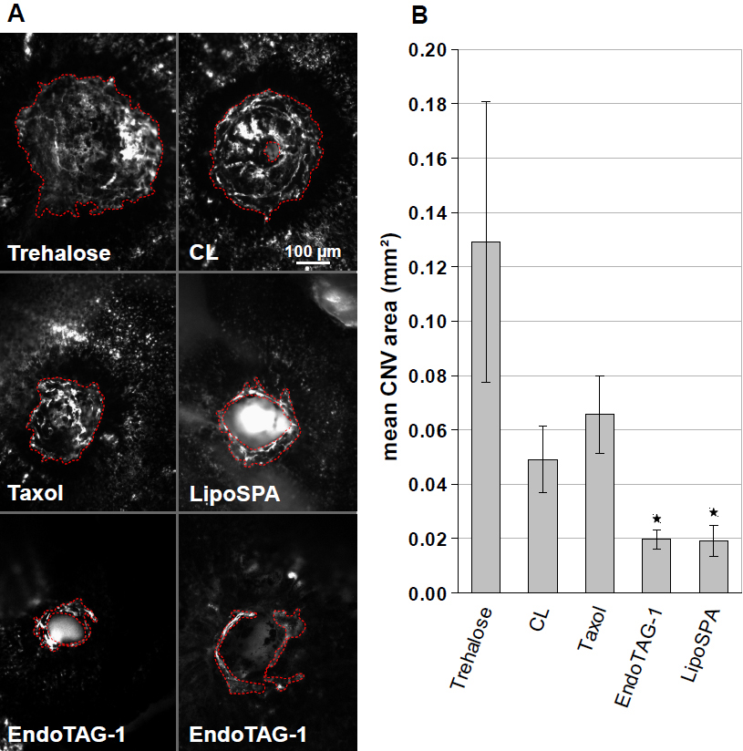
 Figure 2 of
Gross, Mol Vis 2013; 19:54-61.
Figure 2 of
Gross, Mol Vis 2013; 19:54-61.