Figure 2. Suppression of choroidal neovascularization formation by SRPIN340. A: Representative micrographs of choroidal neovascularization (CNV) lesions in the choroidal flat mounts from mice treated
with laser photocoagulation alone as control (CTR, n=32; n represents the number of CNV lesions), combined with intravitreal
injection of 0.1% DMSO (n=31) or 0.2 pmol (n=17), 2 pmol (n=33), and 20 pmol (n=23) SRPIN340, respectively. B: Quantitative analysis of CNV size. Bars indicate the average of CNV size in each group. Values are mean±SEM. *, p<0.05;
†, p<0.01.
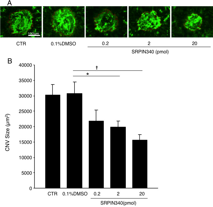
 Figure 2 of
Dong, Mol Vis 2013; 19:536-543.
Figure 2 of
Dong, Mol Vis 2013; 19:536-543.