Figure 4. All images are oriented with the epithelial side up and the stromal side down. Examples of tissue with a small (A, C, E, G) and highnumber of inflammatory cells (B, D, F, H) are shown. Haematoxylin and eosin staining show the morphology of crypts with a small (A) and high (B) number of inflammatory cells. The arrow heads indicate the subepithelial infiltration of the inflammatory cells while the
arrows take the shape of the crypts profile surrounded by inflammatory cells. Inserts: p63 positive cells were evident in
the lower and middle layers of the epithelium in both inflamed tissues (C and D). Few vimentin positive cells were observed in the samples with a high number of inflammatory cells (F) while they were absent in the other one (E). Integrin β4 staining was negative (G and H) in both inflammatory conditions. The images were magnified 200 times.
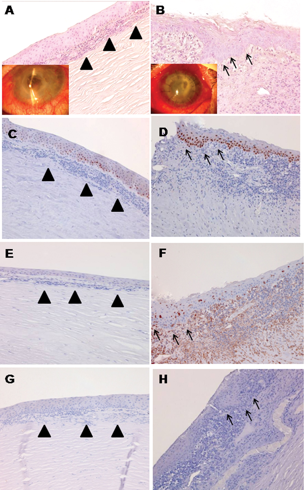
 Figure 4 of
Nubile, Mol Vis 2013; 19:516-525.
Figure 4 of
Nubile, Mol Vis 2013; 19:516-525.