Figure 3. Confocal microscopy images of High (A) and Low (B) density crypts were stained for p63 (green), vimentin (red), and nuclei (blue). Vimentin positive cells were in the basal
layer of crypt (arrow head), and p63 positive cells were in the superficial layer (arrow), which was always negative for vimentin.
The image was magnified 630 times.
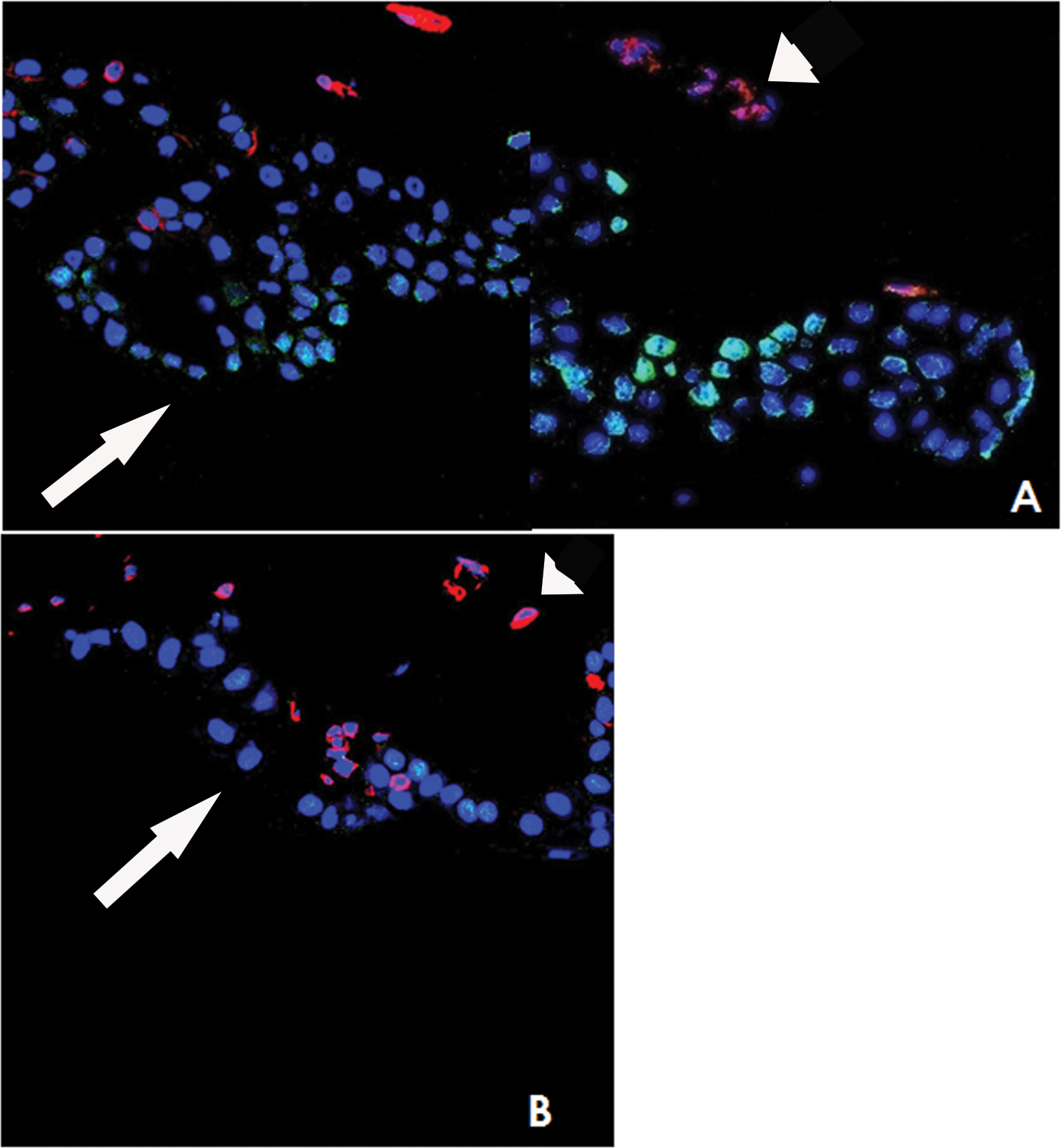
 Figure 3 of
Nubile, Mol Vis 2013; 19:516-525.
Figure 3 of
Nubile, Mol Vis 2013; 19:516-525.