Figure 2. Apoptotic signaling pathways in neuroretina from diabetic patients. A: Protein extracts were prepared from neuroretina from diabetic patients (n=5) and nondiabetic control individuals (n=5).
Total protein (50 μg) was used for western blot analysis with the antibodies against Fas ligand (FasL), caspase-8, BH3 interacting-domain
death agonist (Bid), B-cell lymphoma 2 interacting mediator of cell death (Bim), and active caspase-3. Anti-GADPH antibody
was used as a loading control. B: Autoradiograms were quantified with scanning densitometry. The results are expressed as arbitrary units of protein expression
and are means±SD. *p<0.05, **p<0.01 and ***p<0.005 diabetic patients (D) vsersus control individuals (C).
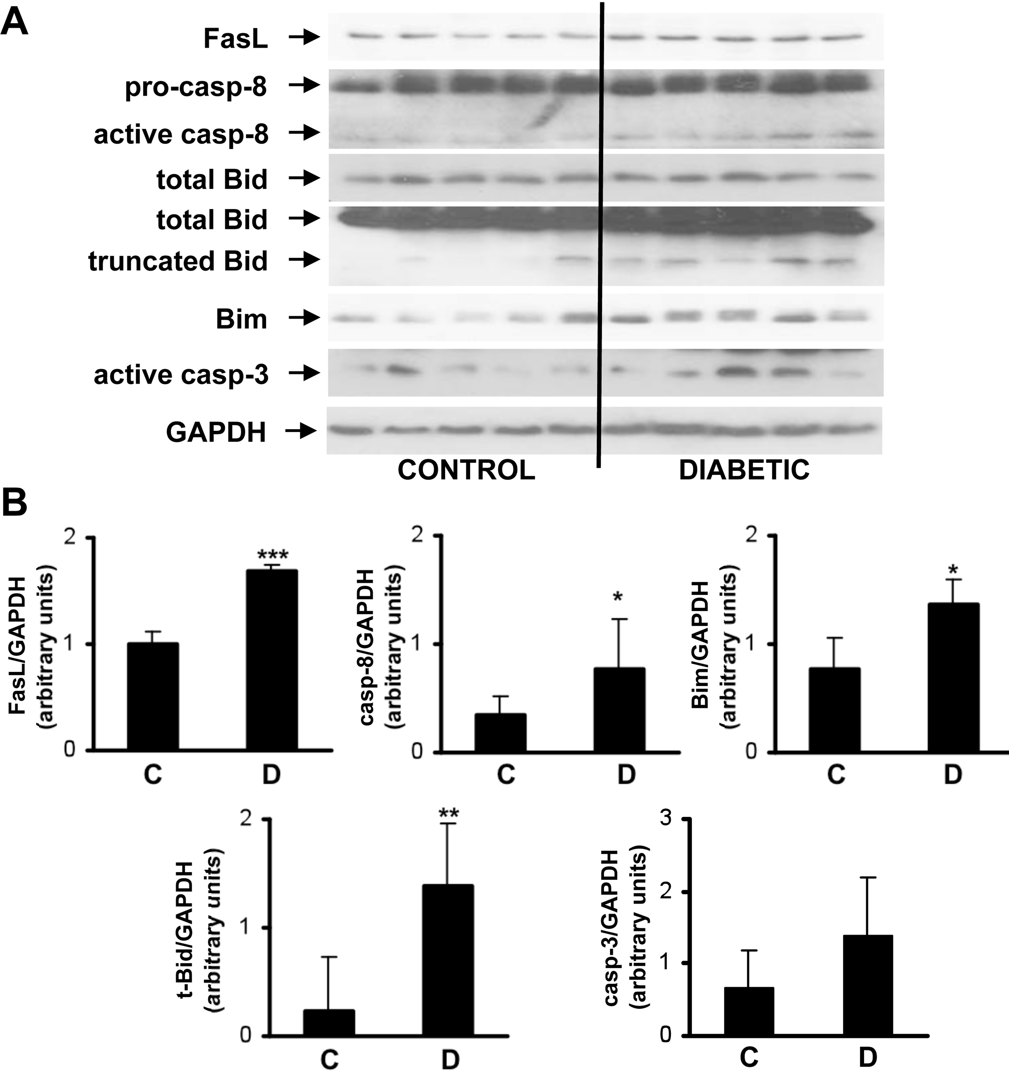
 Figure 2 of
Valverde, Mol Vis 2013; 19:47-53.
Figure 2 of
Valverde, Mol Vis 2013; 19:47-53.