Figure 6. Representative transmission electron microscopy (TEM) photographs of corneas in tissue-engineered human corneal endothelium
(TE-HCE) eye (A, D) normal control eye B, E and denuded amniotic membrane (dAM) eye C from transplanted cats. DM represents Descemet’s membrane. White arrow pointed out the intercellular tight junctions, white
arrow heads pointed out the anchoring junctions such as adherens junctions and desmosomes, black arrow pointed out the gap
junctions, while black arrow heads pointed out the mitochondria. Scale bars A, B, C 2 μm; D, E 200 nm.
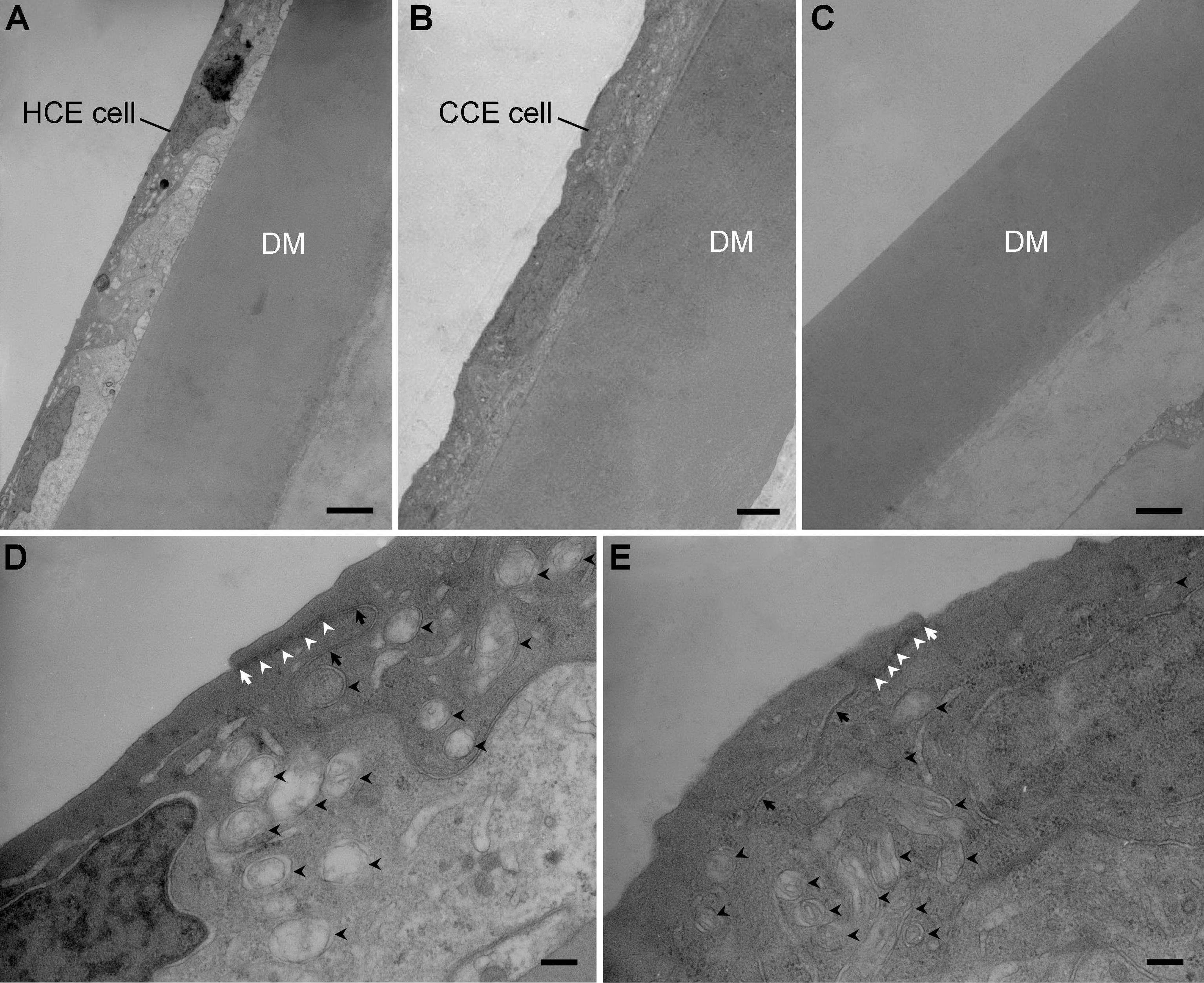
 Figure 6 of
Fan, Mol Vis 2013; 19:400-407.
Figure 6 of
Fan, Mol Vis 2013; 19:400-407.