Figure 5. Histological structures of corneas from transplanted cats. Representative corneal photomicrographs of hematoxylin and eosin
(H&E) staining in tissue-engineered human corneal endothelium (TE-HCE) eye A and normal control eye B SEM in TE-HCE eye C and normal control eye D are shown. Scale bars A, B 100 μm; C, D 10 μm.
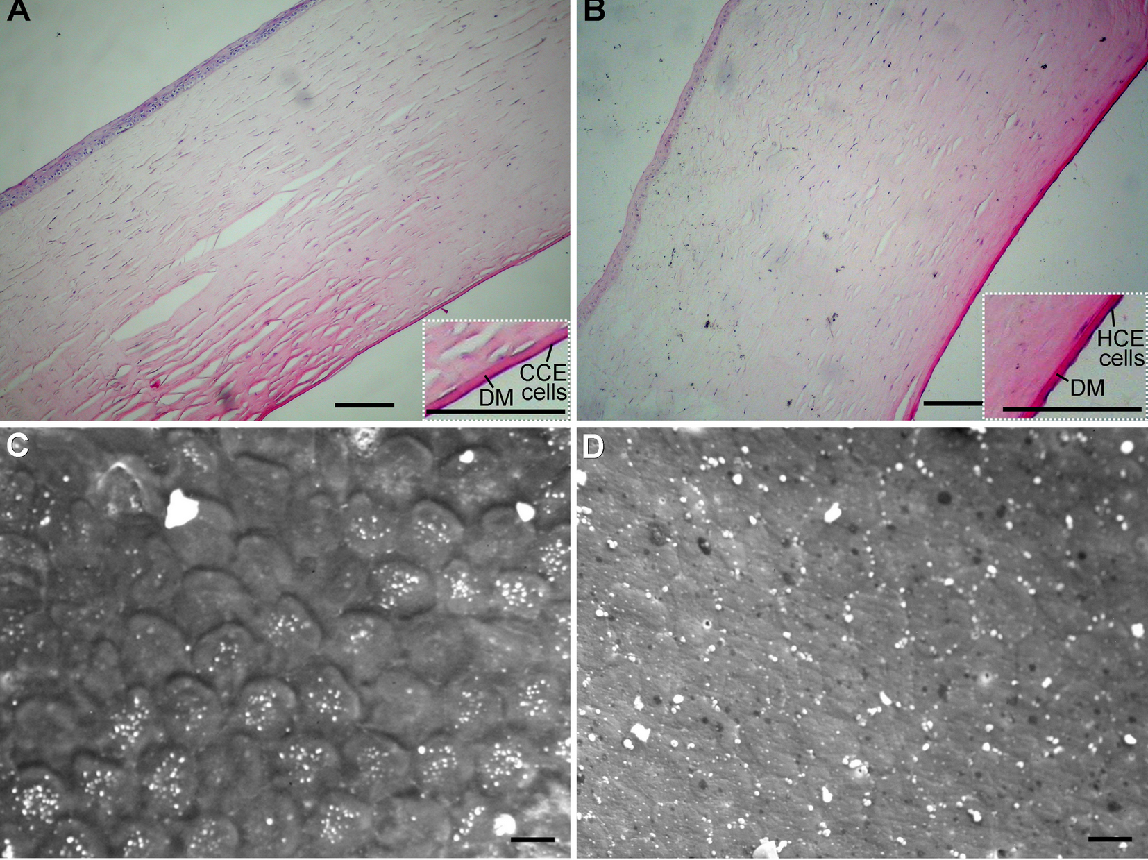
 Figure 5 of
Fan, Mol Vis 2013; 19:400-407.
Figure 5 of
Fan, Mol Vis 2013; 19:400-407.