Figure 4. Corneal endothelia from transplanted cats. Fluorescent photographs of 1,1'-dioctadecyl-3,3,3',3'-tetramethylindocarbocyanine
perchlorate (DiI)-labeled monoclonal human corneal endothelium (HCE) cells in tissue-engineered (TE)-HCE eye A and denuded amniotic membrane (dAM) eye B alizarin red staining photographs of the monoclonal HCE cells in TE-HCE eye C and normal control eye D are shown. In the TE-HCE eye A, a cell-visible high magnification view of the cornea is shown in the box with a dashed line. Scale bars A, B 100 μm; C, D 50 μm.
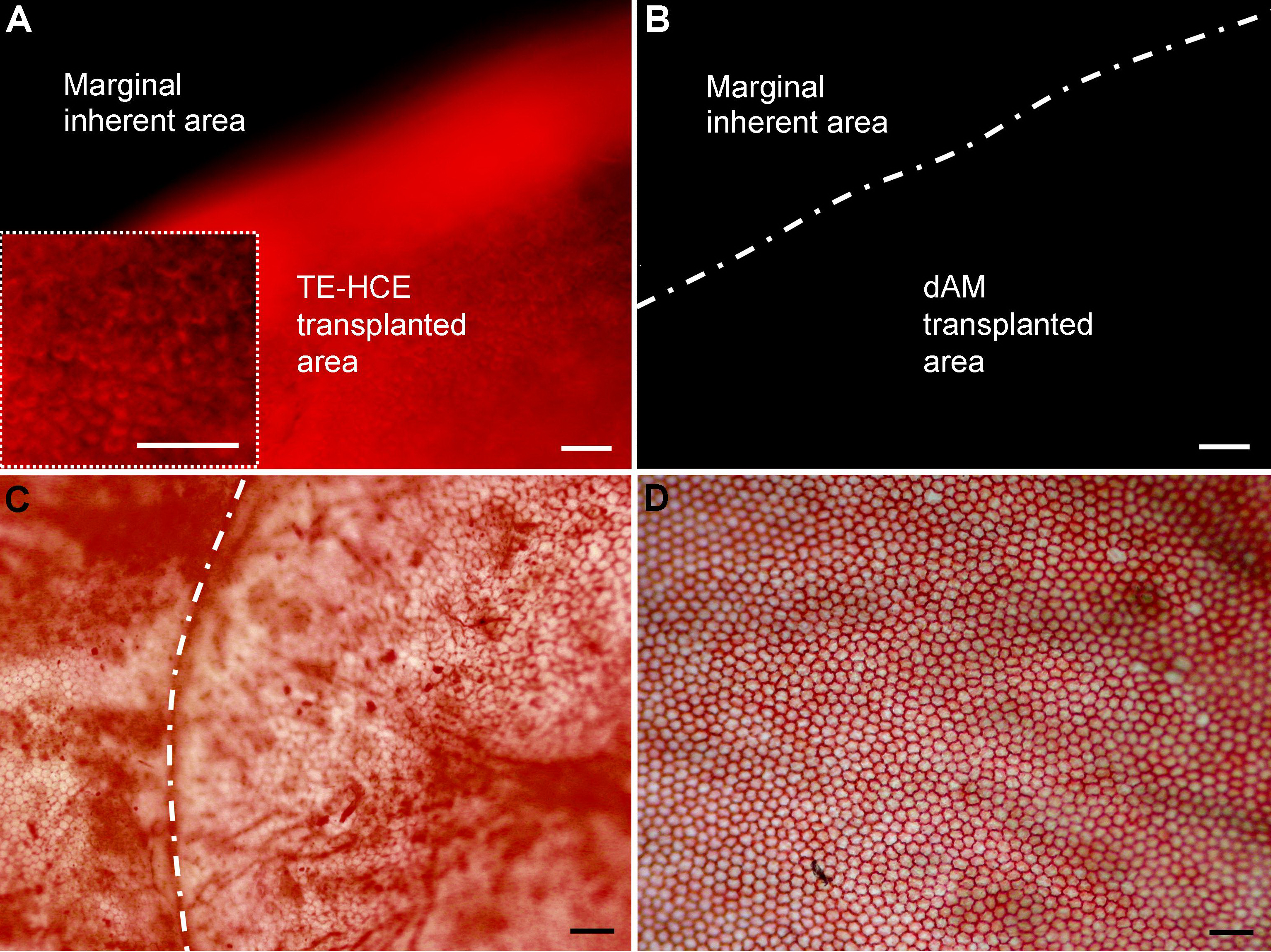
 Figure 4 of
Fan, Mol Vis 2013; 19:400-407.
Figure 4 of
Fan, Mol Vis 2013; 19:400-407.