Figure 1. The tissue-engineered human corneal endothelium reconstructed from monoclonal human corneal endothelium (HCE) cells and denuded
amniotic membrane (dAM) at day 4. The morphology and structure of tissue-engineered (TE)-HCE cells are shown in 1% alizarin
red staining A. Frozen sections were stained with hematoxylin and eosin (H&E), B and visualized using scanning electron microscopy (SEM) C and transmission electron microscopy (TEM) D. Scale bars, A 20 μm; B 100 μm; C 10 μm; D 2 μm.
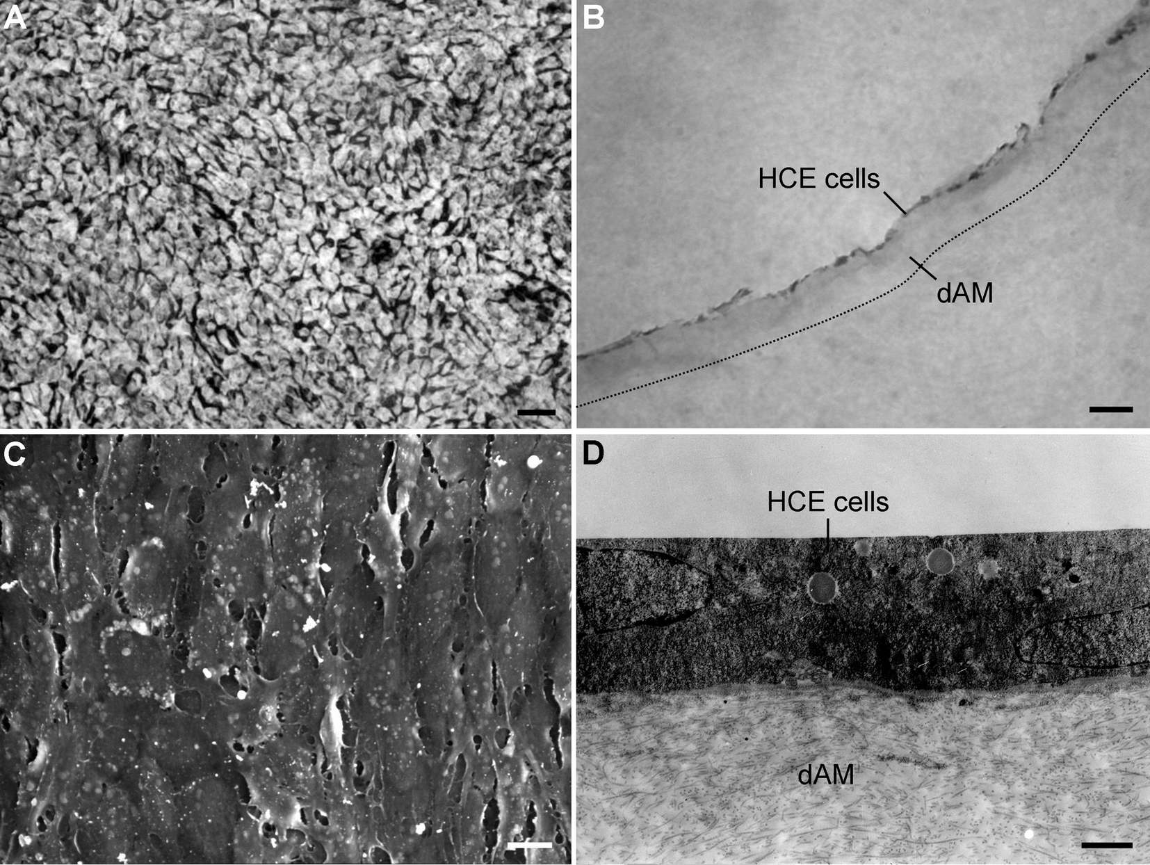
 Figure 1 of
Fan, Mol Vis 2013; 19:400-407.
Figure 1 of
Fan, Mol Vis 2013; 19:400-407.