Figure 4. P53 expression in human pterygium. Basal and parabasal distribution of p53 positive cells in the human pterygium (A, B). Scattered positive p53 positive fibroblast-like cells in fibrovascular compartment of human pterygium (C). Note the discrepancies between the higher density of the p53 positive cells in the basal layer of the epithelial compartment
and the lack of p53 expression in the stromal component. Blood and lymphatic endothelial cells positive for p53, with high
intensity for blood vessels (D) and low density for lymphatics (E).
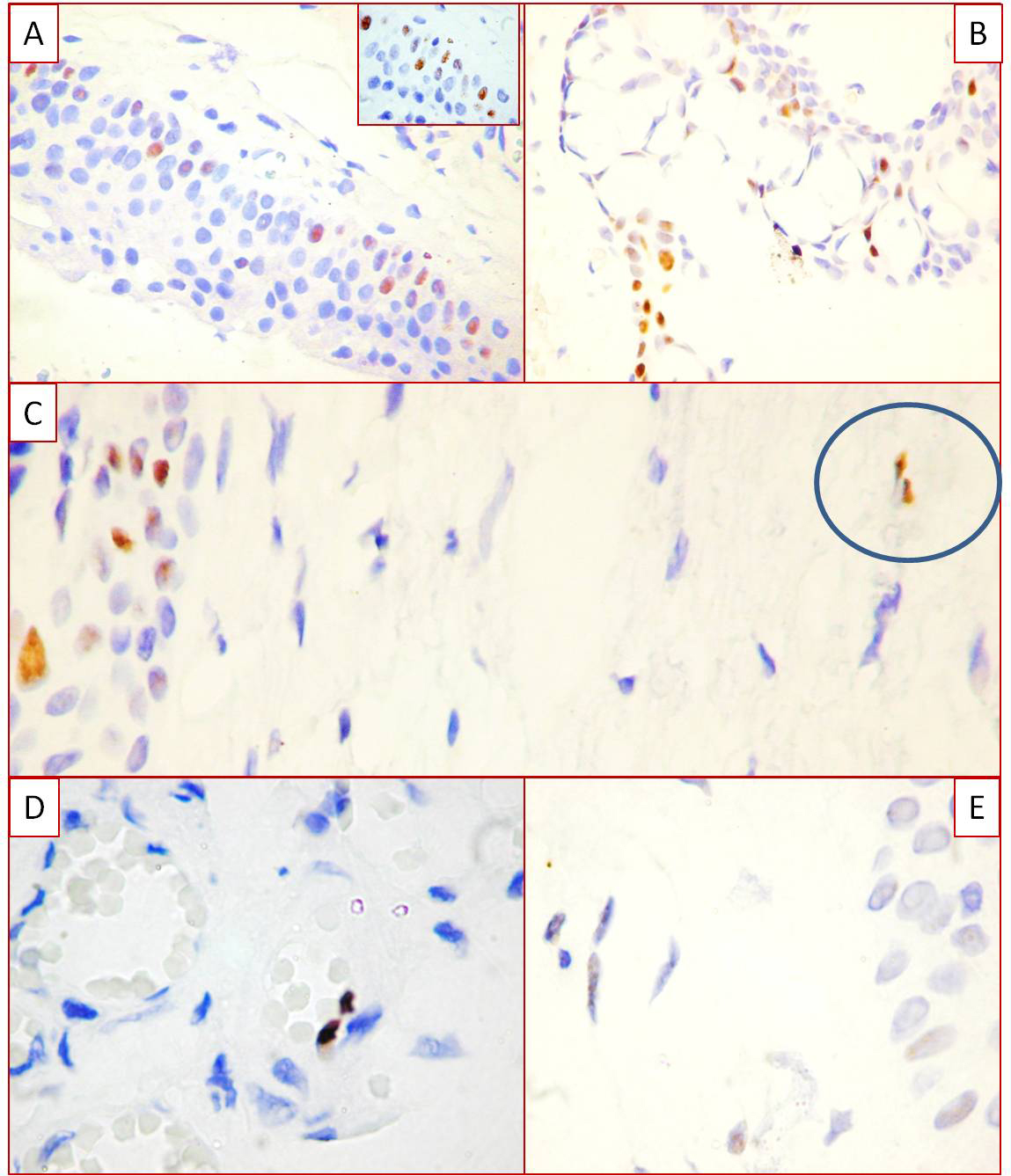
 Figure 4 of
Cimpean, Mol Vis 2013; 19:348-356.
Figure 4 of
Cimpean, Mol Vis 2013; 19:348-356.