Figure 3. Thymine dimer expression in the fibrovascular compartment of human pterygium. Epithelial and fixed connective tissue cells
were positive for thymine dimer reaction (A). Small blood vessels’ endothelial cells constantly had nuclear expression in almost all endothelial areas (B). In contrast, for mature blood vessels’ endothelial cells from preexisting vessels no immunohistochemical expression of
DNA damage was found (C). Endothelial cells from the tip of the vascular sprout intensely stained for thymine dimers (D). Covering pericytes had a moderate intensity of thymine dimer expression compared with the capillary endothelial cells (E). Thymine dimers expressing endothelial cells line the lymphatic vessel lumen (F).
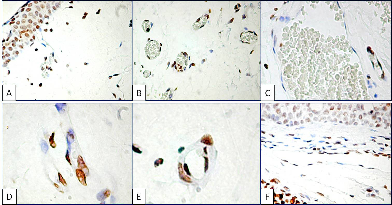
 Figure 3 of
Cimpean, Mol Vis 2013; 19:348-356.
Figure 3 of
Cimpean, Mol Vis 2013; 19:348-356.