Figure 7. Transmission electron microscope images of skin tissue sections from patients with p.Tyr1792fsX55-causing mutations and age
matched control individuals. Representative electron micrographs of a homozygous carrier with primary congenital glaucoma
(PCG; A) and the control individual (C), and a heterozygous carrier with pseudoexfoliation (PEX) syndrome (B) and the control individual (D). More areas that appear devoid of extra cellular matrix (ECM) structures are evident in the image taken from the patients’
samples (A, B), and the ECM of the patient with PCG (A) is sparser than that of his older mother with PEX syndrome (B). Examples of sparse regions are shown with a star (∗). f, fibroblast; cb, collagen bundles.
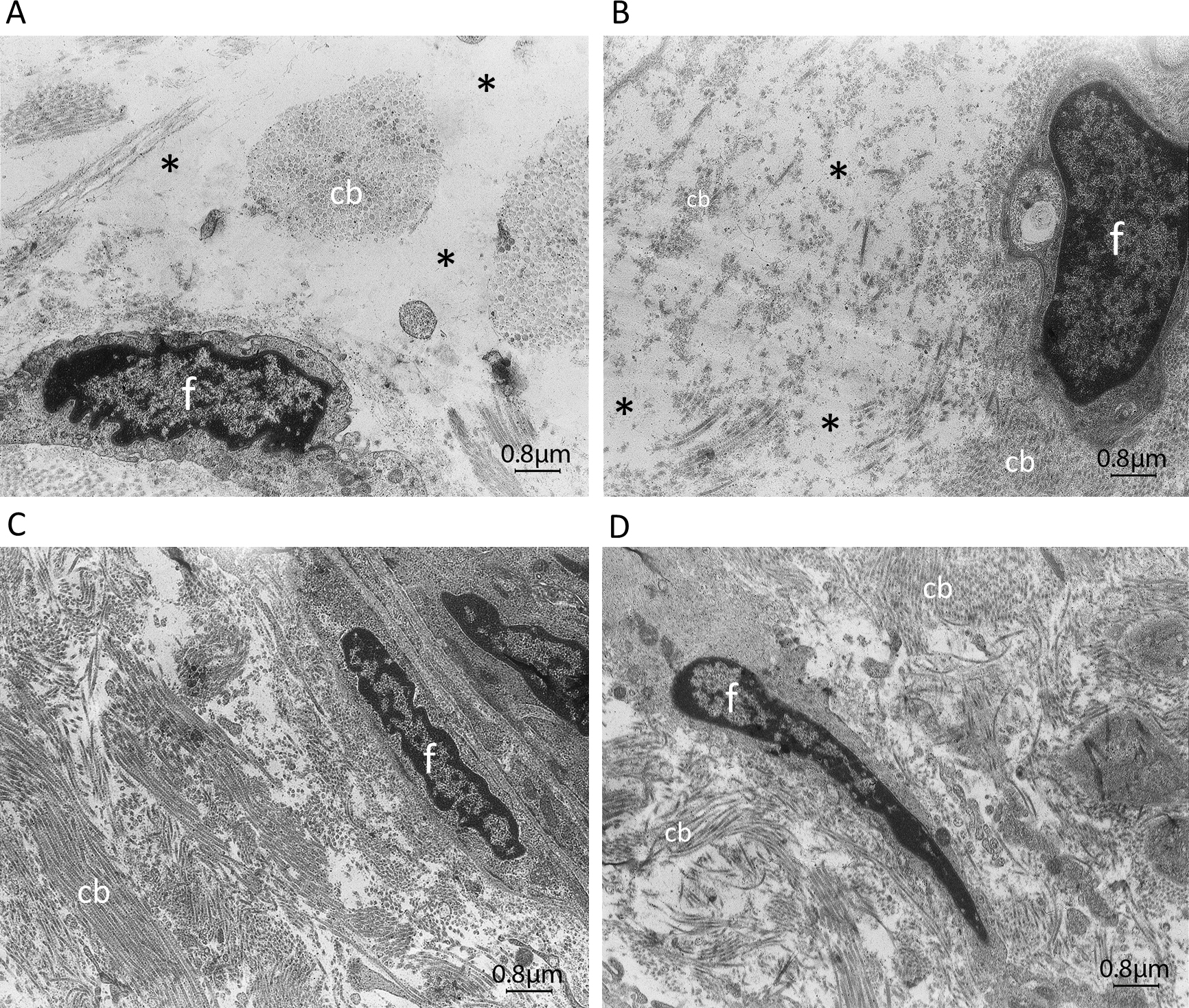
 Figure 7 of
Jelodari-Mamaghani, Mol Vis 2013; 19:333-347.
Figure 7 of
Jelodari-Mamaghani, Mol Vis 2013; 19:333-347.