Figure 6. Light microscope images of skin tissue sections from patients with p.Tyr1792fsX55-causing mutations and age-matched control
individuals. A–D: Orcein Geimsa–stained elastic fibers of a homozygous carrier with primary congenital glaucoma (PCG; A) and control individual (C), a heterozygous carrier with pseudoexfoliation (PEX) syndrome (B) and a control individual (D) visualized with a light microscope. Elastic fibers were sparser and fragmented in each patient compared to the respective
control. Examples of fragmented fibers and sparse regions are shown, respectively, with arrows and * symbol. E–H: Trichrome stained collagen fibers of individual with PCG (E) and control individual (G), individual with PEX syndrome (F) and control individual (H) visualized with a light microscope.
Collagen fibers, stained with blue, were sparser in each patient compared to the respective control. Arrector pili muscles
appear red in G.
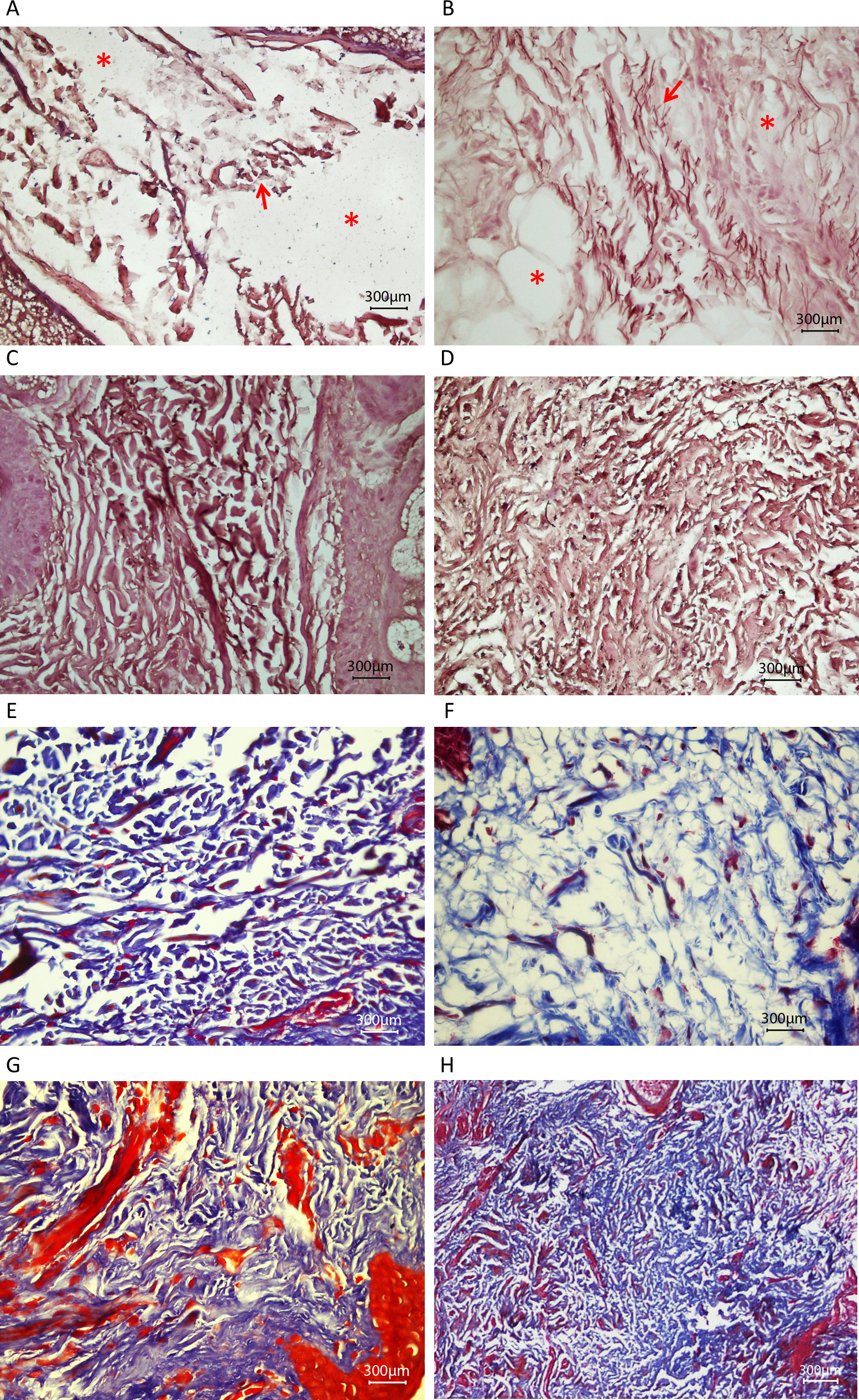
 Figure 6 of
Jelodari-Mamaghani, Mol Vis 2013; 19:333-347.
Figure 6 of
Jelodari-Mamaghani, Mol Vis 2013; 19:333-347.