Figure 5 of
Jelodari-Mamaghani, Mol Vis 2013; 19:333-347.
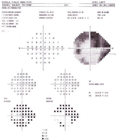
Figure 5.
Visual field defects in the left eye of patient with POAG (211L). The single field analysis printout shows severe visual field loss with dense superior arcuate scotoma that threatens the fixation and inferior nasal field defect.
 Figure 5 of
Jelodari-Mamaghani, Mol Vis 2013; 19:333-347.
Figure 5 of
Jelodari-Mamaghani, Mol Vis 2013; 19:333-347.