Figure 10. Images of eyes of a patient with pseudoexfoliation glaucoma syndrome and a patient with primary congenital glaucoma with heterozygous
and homozygous p.Tyr1792fsX55-causing mutation in LTBP2, respectively. A and B: Images showing deposition of pseudoexfoliation material (arrows) on the endothelial surface of the cornea (arrow in A) and at the pupillary border of the iris (arrow in B) in the eye of an individual diagnosed with pseudoexfoliation glaucoma (PEXG) syndrome. C: Fundus photograph of the same PEXG individual exhibiting characteristic features of glaucomatous optic neuropathy. Diffuse
neuroretinal rim thinning with more involvement of the inferior rim, notching and peripapillary atrophy all around the disc
are evident. D: Image of an eye of an individual with PCG showing diffuse corneal opacity and calcific band keratopathy.
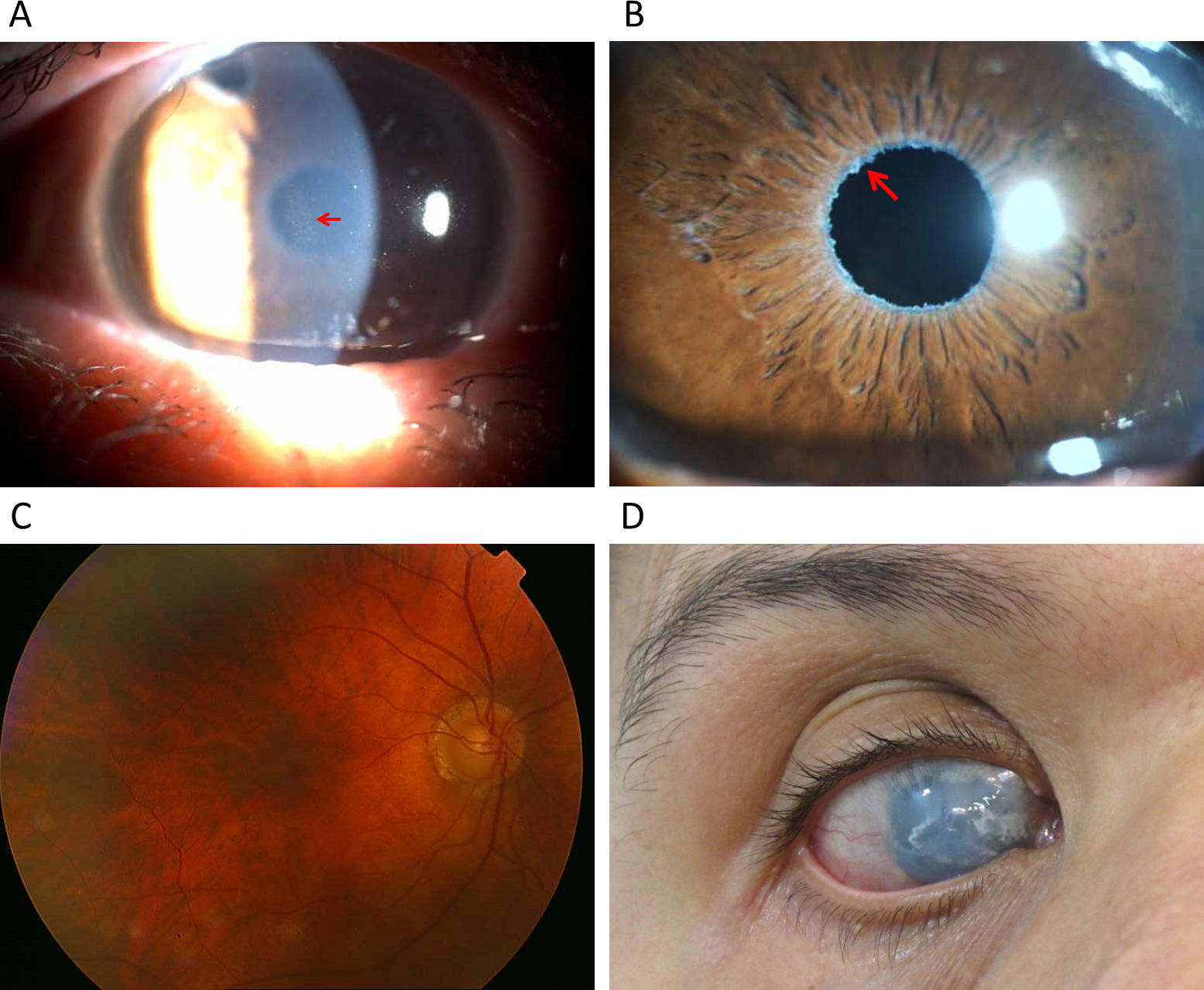
 Figure 10 of
Jelodari-Mamaghani, Mol Vis 2013; 19:333-347.
Figure 10 of
Jelodari-Mamaghani, Mol Vis 2013; 19:333-347.