Figure 4. Cell-cell junction formation in the ShCDK5 human corneal limbal epithelial cells. Human corneal limbal epithelial (HCLE) cells
(A, B), in the absence of cyclin-dependent kinase 5 (CDK5) with olomoucine (C, D) or ShCDK5 (E, F) and lens epithelial cells (NN1003; G, H) were treated with olomoucine (I, J). The cells were allowed to form cell–cell adhesions in the presence or absence of calcium for 1 h at 37 °C, and particle
aggregates were cytospin on to glass slides to measure the size of the aggregates. Calcium-dependent E-cadherin junctions
were formed in the presence of CDK5 (B, K) in the HCLE cells in contrast to N-cadherin expressing lens epithelial cells that form larger aggregates in the absence
of CDK5 (J, K). (A–J) 20X images. E-cadherin and N-cadherin induction during cell–cell junction formation in the MDA-MB-231 cells (L). Green fluorescent protein (GFP)-E-cadherin and GFP-N-cadherin transfected cells were allowed to form cell–cell aggregates
in the absence of calcium (lane 1), presence (lane 2) and with olomoucine (lane 3). Cells containing E-cadherin form more
cell–cell aggregates (group 3) while the N-cadherin (group 4) aggregates increases in the presence of olomoucine. N-cadherin
induction in the ShCDK5 and olomoucine HCLE (M) CDK5 inhibition leading to degradation of E-cadherin leads to induction of N-cadherin. P120 coimmuneprecipitates with N-cadherin
to stabilize the junctional complex in the ShCDK5 and olomoucine-treated HCLE. IP=immunoprecipitation; WCl=whole cell lysate.
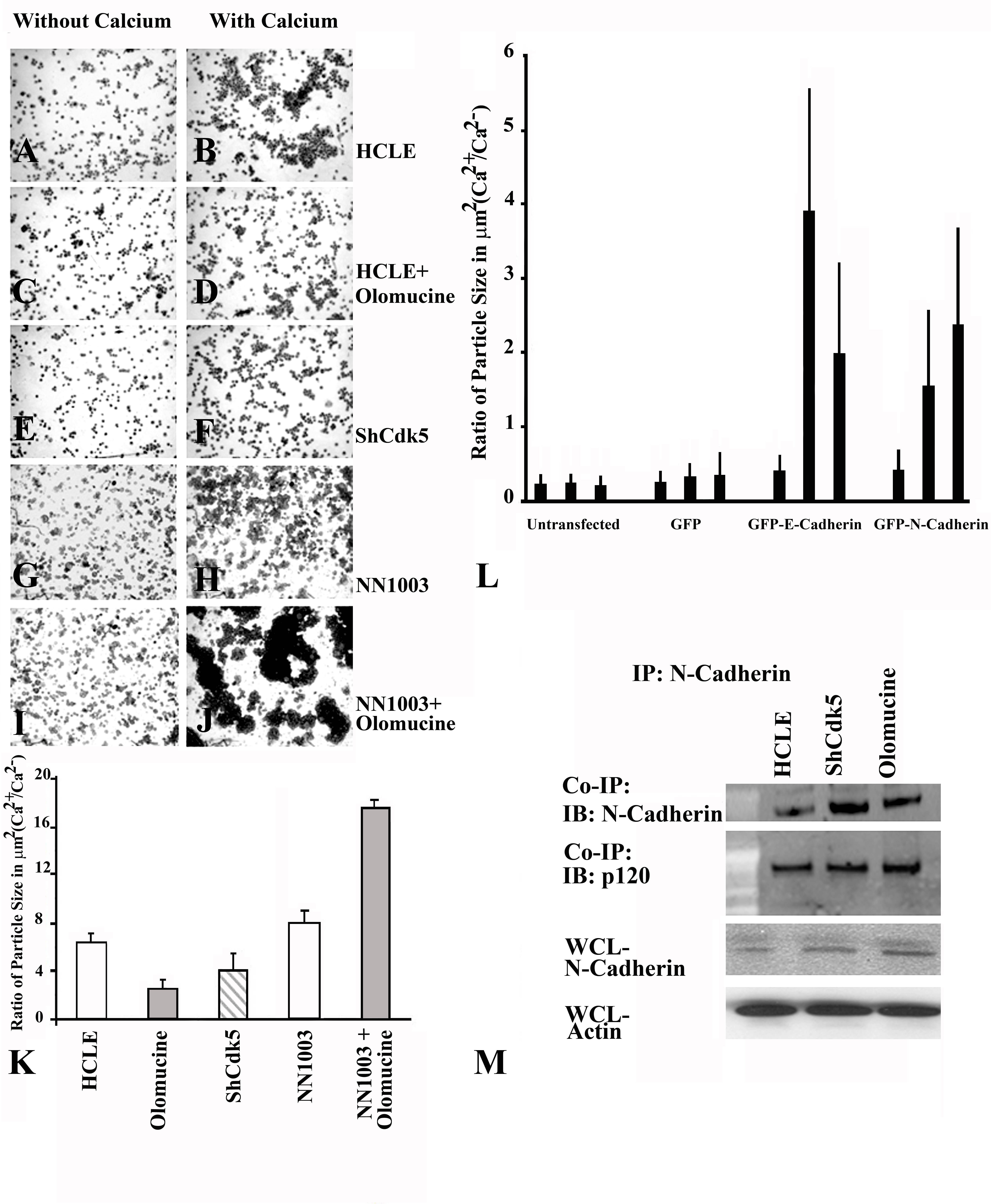
 Figure 4 of
Arpitha, Mol Vis 2013; 19:319-332.
Figure 4 of
Arpitha, Mol Vis 2013; 19:319-332.