Figure 2. Human corneal limbal epithelial cell line suppressed for cyclin-dependent kinase 5 expression. Immunofluorescence image showing
staining for cyclin-dependent kinase 5 (CDK5) (green; A) in human corneal limbal epithelial (HCLE) cells and note the absence of Cdk5 in the ShCDK5 cells (C). Nuclear counterstaining 4', 6-diamidino-2-phenylindole (blue) for images in A and C (B, D). Lysates from these cells showing significant reduction in the expression levels of CDK5 in the plentiviral transduced HCLE
(ShCDK5) cells (E). 100X images.
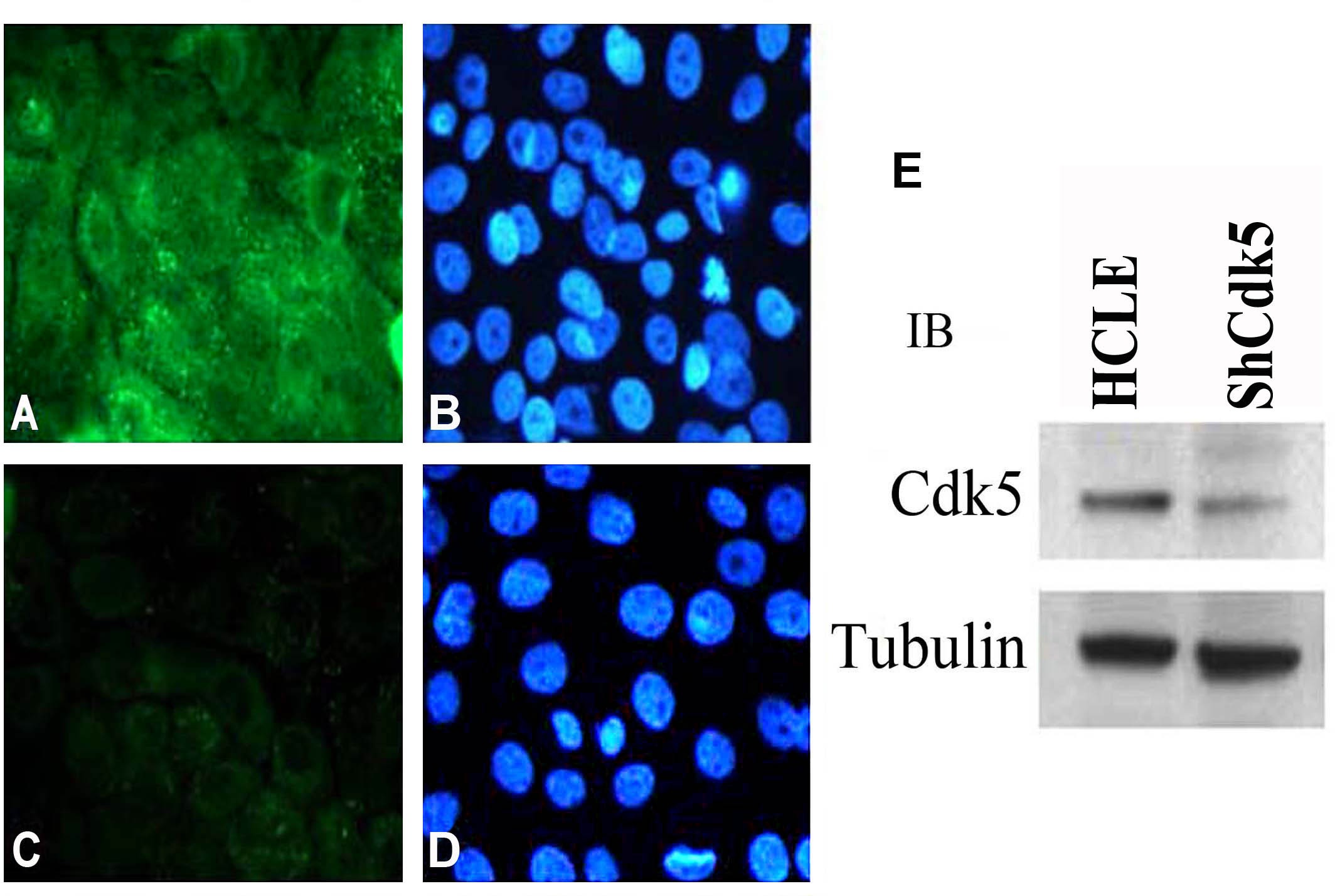
 Figure 2 of
Arpitha, Mol Vis 2013; 19:319-332.
Figure 2 of
Arpitha, Mol Vis 2013; 19:319-332.