Figure 1. Co-localization of E-cadherin and cyclin dependent kinase5 (Cdk5) in the cell–cell borders. Human corneal limbal epithelial
(HCLE) cells were grown on chamber slides, fixed, and immunostained stained with pY15 (green; A) and E-cadherin (red; B). Cyclin-dependent kinase 5 (Cdk5) and E-cadherin were localized to the cell–cell boundaries, and the overlay image demonstrates
the colocalization (C) of E-cadherin and phosphorylated (pY15) CDK5. Confluent cultures of HCLE showing CDK5 border localization (D) confluent HCLE cultures when treated with olomoucine show a shift in the localization of pY15 CDK5 from the cell borders
to the interior (E). E-cadherin and CDK5 form a part of the same protein complex (F, G) E-cadherin coimmuneprecipitates with CDK5 (38 kDa; F). Affinity chromatography for HCLE cell lysates were pulled down for
glutathione-S-transferase-cyclin-dependent kinase 5 (GST-CDK5) and that glutathione beads bound to CDK5 formed a complex with
E-cadherin (G). (A, B, C) 100X images and (D, E) 40X images.
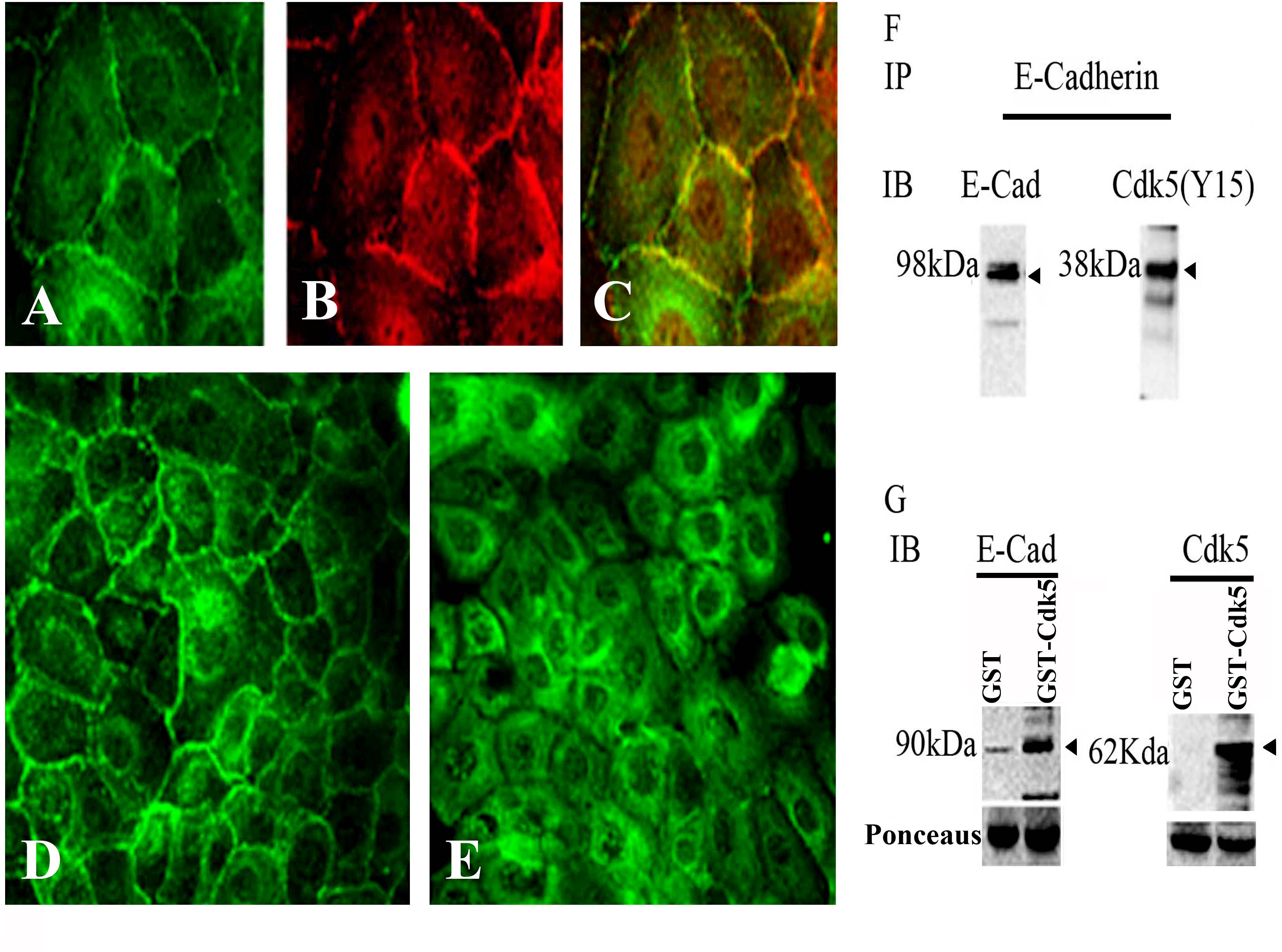
 Figure 1 of
Arpitha, Mol Vis 2013; 19:319-332.
Figure 1 of
Arpitha, Mol Vis 2013; 19:319-332.