Figure 3. All images from the mother of the proband. A: Multiple erythematous scars in neck (two indicated by arrows); lesions had aspect of hemorrhaging incisions in the neonatal
period. B and C: Fundus abnormalities with white, drusenoid deposits and reticular RPE abnormalities in the macula (B: Small and large arrow respectively), and whole fundus of left eye (C), with reticular abnormalities with alternating hypo- and hyperpigmentation of retinal pigment epithelium (RPE). D: Hyper- and hypointense areas on fluorescein angiography in keeping with RPE abnormalities described under A. E: Full-field flash electroretinography of the right eye (top traces); traces at bottom are from normal age-matched control
for purposes of comparison. Notice overall reduction of retinal function for rod-specific, combined rod-cone, and cone-specific
responses to about three-fifths of normal amplitudes, without significant delay in responses. Retinal function is thus reduced
in keeping with the reduced total surface area of the retina, but the remaining areas function normally, suggesting this is
not progressive retinal degeneration.
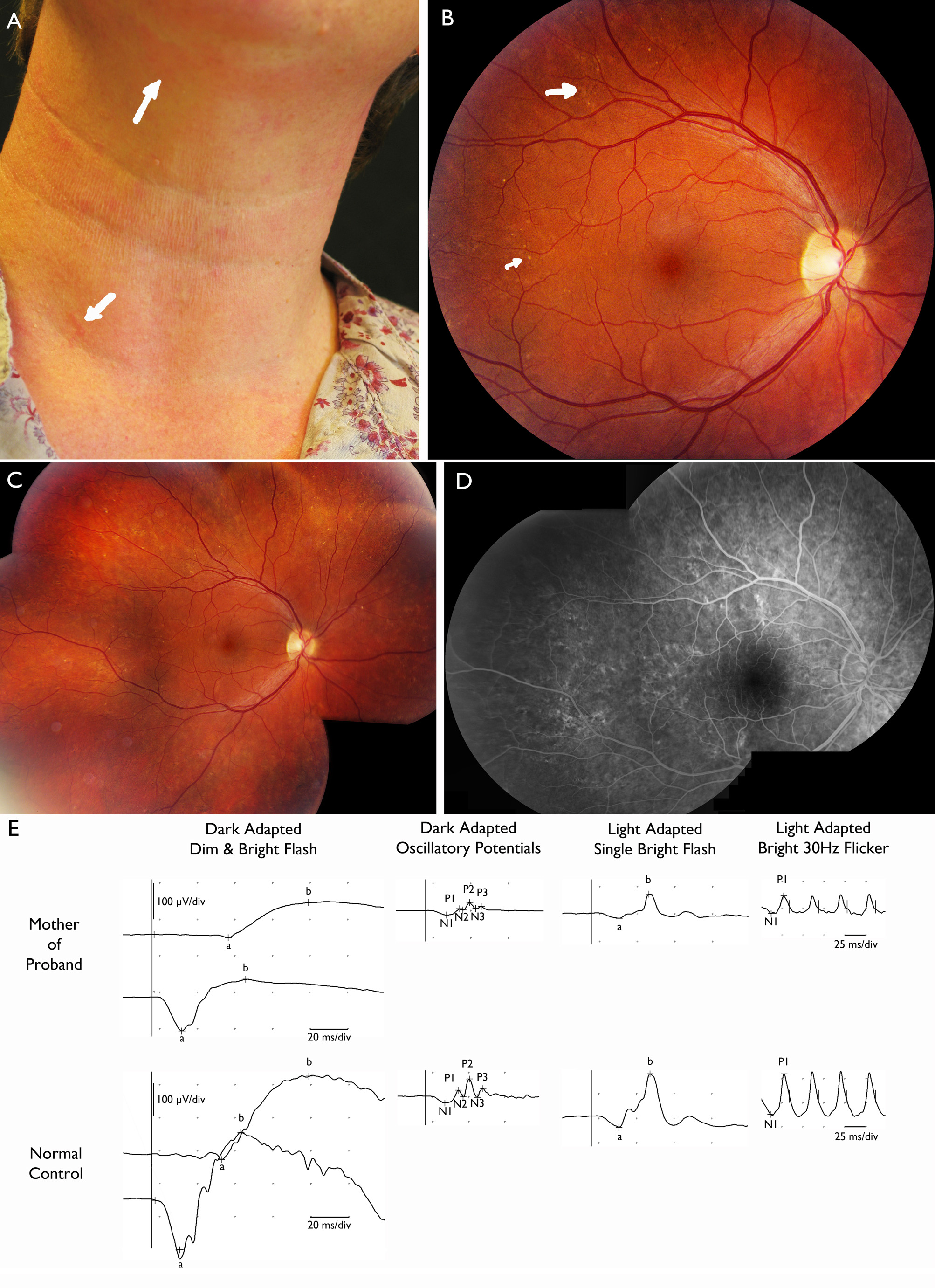
 Figure 3 of
Vergult, Mol Vis 2013; 19:311-318.
Figure 3 of
Vergult, Mol Vis 2013; 19:311-318.