Figure 2. All images from the proband. A: Microphthalmic right eye and clinically normal left eye; notice esotropia and corneal opacification of the right eye. B: Right eye with corneal opacification in central and inferotemporal areas. C: Detail of inferotemporal area of right eye; notice patent peripheral corneal vessels (arrow) in area of reticular endothelial
abnormalities and relative stromal opacification. D: Detail of central area of right eye; notice central corneal ghost vessels (arrow) in area of corneal opacification.
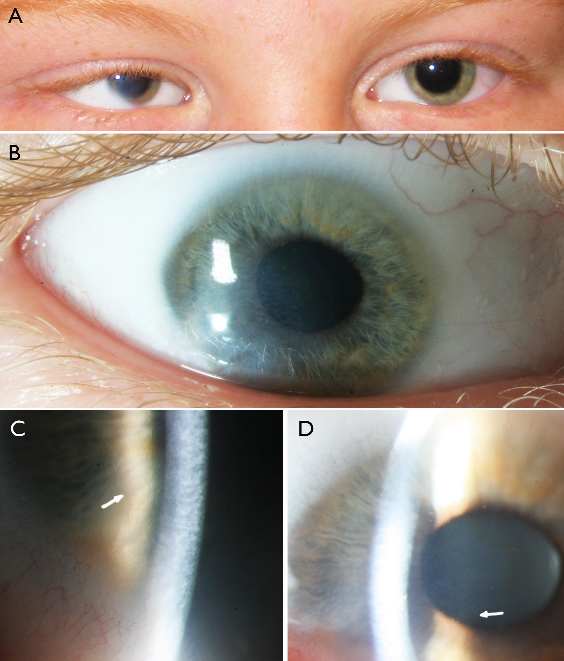
 Figure 2 of
Vergult, Mol Vis 2013; 19:311-318.
Figure 2 of
Vergult, Mol Vis 2013; 19:311-318.