Figure 4. Induction of the signaling pathway in the presence of interleukin 1 beta (IL-1β), tumor necrosis factor alpha (TNF-α), and
interferon gamma (IFN-γ). In ARPE-19 cells, cell lysates were analyzed with western blot for phosphorylated MAP kinase signaling
protein (extracellular-signal-regulated kinases [ERK1/2], p38, and c-Jun N-terminal kinase [JNK]) and IκB and total mitogen
activating protein (MAP) kinase signaling protein and IκB after IL-1β (10 ng/ml; A), TNF-α (10 ng/ml; B), or IFN-γ (10 ng/ml; C) administration. Glyceraldehyde 3-phosphate dehydrogenase (GAPDH) was used as a loading control. Immunoblot analyses of ERK1/2,
p-ERK1/2, p38, p-p38, JNK, p-JNK, IκB, and p-IκB and densitometric quantification (bottom) with or without IL-1β, TNF-α, or
IFN-γ, as indicated. Results are representative for three independent experiments. *p<0.05 versus vehicle.
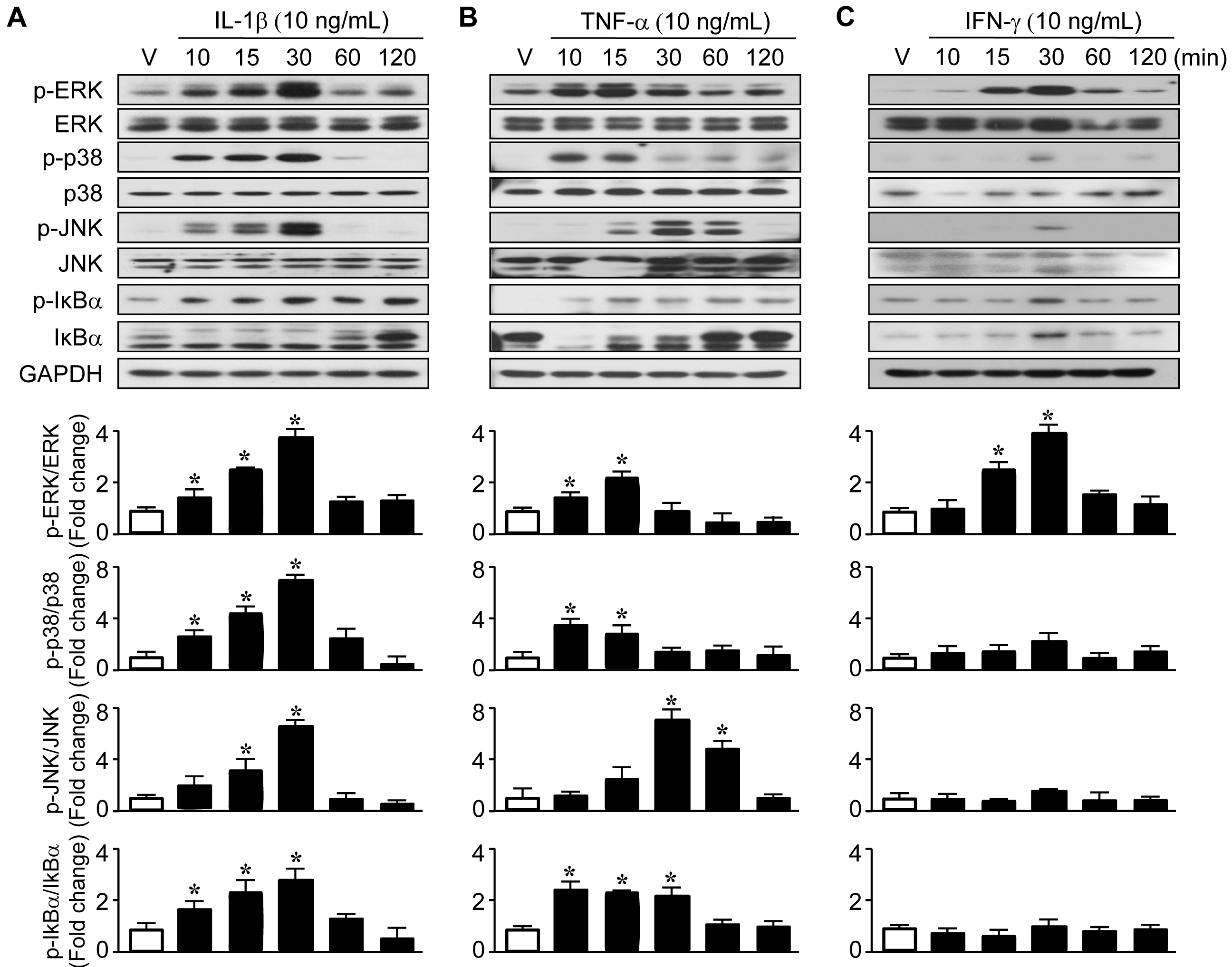
 Figure 4 of
Woo, Mol Vis 2013; 19:303-310.
Figure 4 of
Woo, Mol Vis 2013; 19:303-310.