Figure 4. Vascular endothelial growth factor secretion, transcription factors, and p38. Organ cultures at day 2 of preparation were
treated with indicated inhibitors, and supernatant was collected for 1 h after 6 h, 24 h, and 48 h. Untreated cultures served
as controls. Vascular endothelial growth factor (VEGF) content was evaluated with enzyme-linked immunosorbent assay. Inhibition
of HIF-1α with YC-1 or of Stat3 with Stattic displayed no significant effect on VEGF secretion (A, B). The SP-1 inhibitor mithramycin significantly reduced VEGF secretion after 24 h and 48 h (C). The inhibition of nuclear factor kappaB (NFkB) and of p38 with SB203580 significantly reduced VEGF secretion at all time
points tested (D, E). The results are depicted as % VEGF at 0 h (before treatment). The bars depict the mean and standard deviation of three
to five independent experiments. Absolute concentration of VEGF at 0 h was 202.03±59.04 pg/ml for mithramycin, 202.68±69.16
pg/ml for the NFkB inhibitor, 235.51±52.31 pg/ml for Stattic, 186.23±13.46 pg/ml for YC-1, and 148.58± 93.84 pg/ml for SB203580.
The absolute concentration of VEGF of the respective controls at 0 h was 245.73±78.28 pg/ml for mithramycin, 243.27±22.77
pg/ml for the NFkB inhibitor, 158.43±139.4 pg/ml for YC-1, and 113.90±42.64 pg/ml for p38. Statistical significance was determined
with the Student t test. Significant is depicted as follows: + p<0.05; ++ p<0.01. +++ p<0.001.
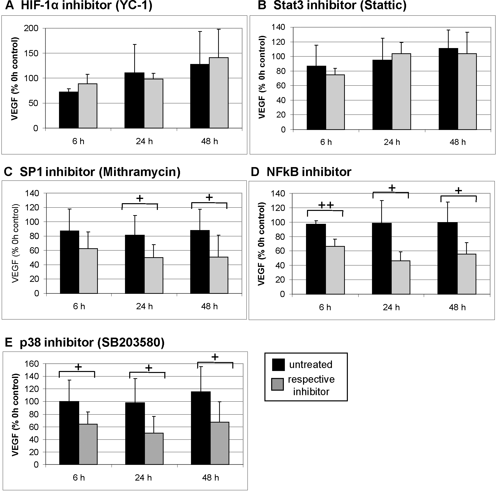
 Figure 4 of
Klettner, Mol Vis 2013; 19:281-291.
Figure 4 of
Klettner, Mol Vis 2013; 19:281-291.