Figure 1. Toxicity of applied biochemical agents. Primary retinal pigment epithelium cells were treated with (A) indicated inhibitors and (B) a combination of inhibitors for 24 h or 48 h and assessed for cell viability in an MTT assay. Untreated cells served as
control. None of the inhibitors displayed a significant toxicity applied concentrations: SU1498: 10 μM; Bisindolylmaleimide:
1 μM; LY294002: 25 μM; NFkB inhibitor: 1 μM; Mithramycin 1 μM; Stattic 1 μM; YC-1: 25 μM; SB203580: 10 μM. Abbreviations:
SU=SU1498; LY=LY294002; Bis: bisindolylmaleimide; NFkB: NFkB inhibitor; SB=SB203580. The bars depict the mean and standard
deviation of three to five independent experiments. Statistical significance was determined with the Student t test.
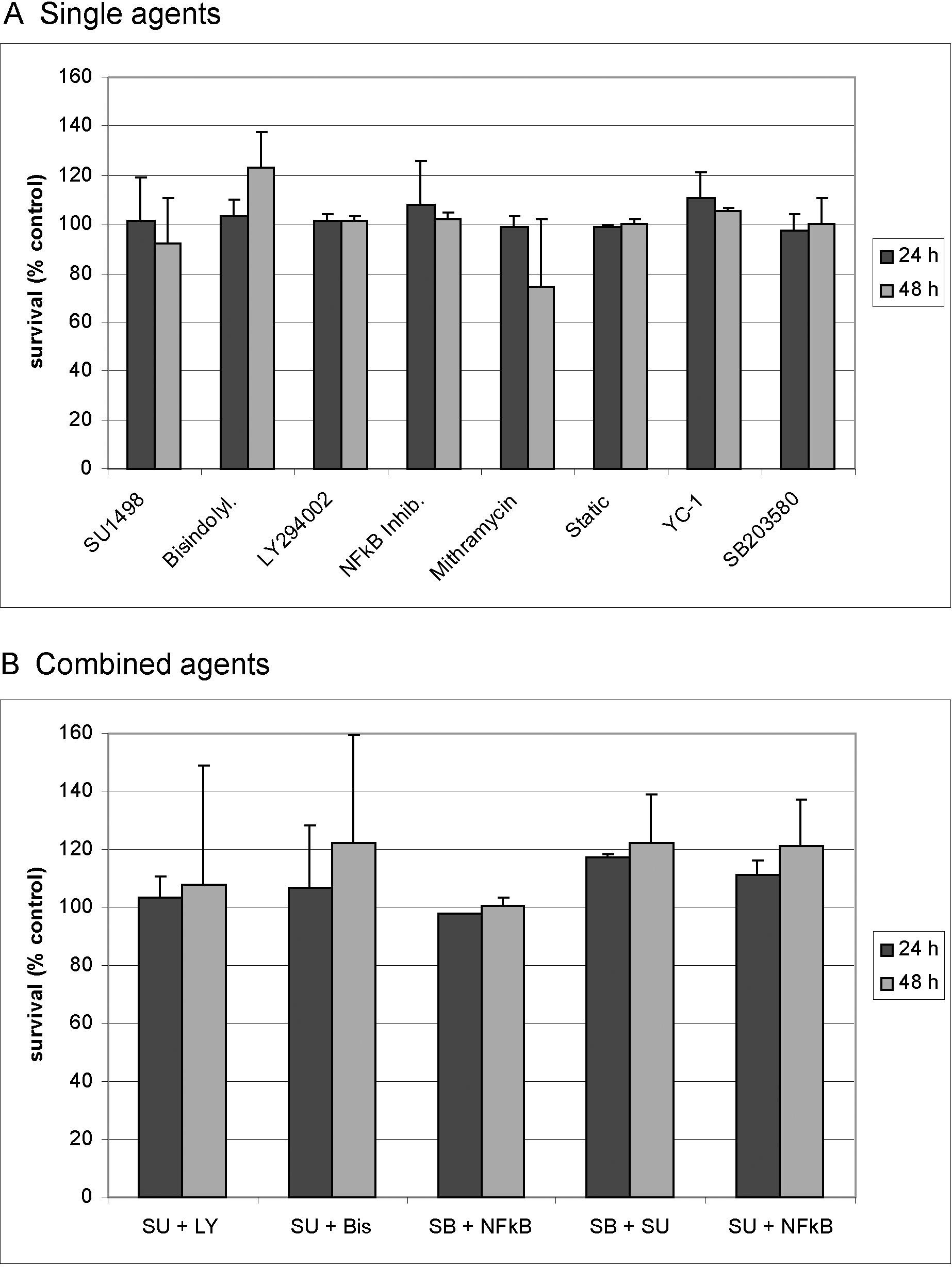
 Figure 1 of
Klettner, Mol Vis 2013; 19:281-291.
Figure 1 of
Klettner, Mol Vis 2013; 19:281-291.