Figure 6. Pirfenidone (PFD) had no effect on cell apoptosis. Retinal pigment epithelial (RPE) cells were incubated for 24 h with complete
medium (control), 0.3 mg/ml pirfenidone, 0.5 mg/ml PFD, or 1.0 mg/ml PFD. A: Cells labeled with Annexin V (-) PI (+) were shown in Q1 area; cells labeled with Annexin V (+) PI (+) were shown in Q2
area; cells labeled with Annexin V (-) PI (-) were shown in Q3 area; cells labeled with Annexin V (+) PI (-) were shown in
Q4 area. B: The percentages of cells labeled as Annexin V (+) PI (-) and Annexin V (+) PI (+) were investigated using flow cytometry.
There were no significant differences between the groups (n=4, the error bar indicates standard deviation [SD], p>0.05).
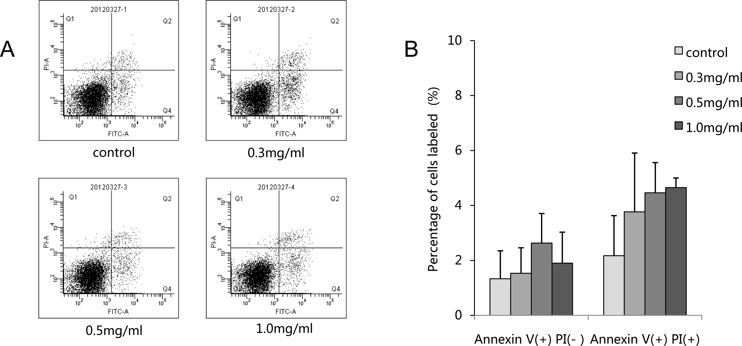
 Figure 6 of
Wang, Mol Vis 2013; 19:2626-2635.
Figure 6 of
Wang, Mol Vis 2013; 19:2626-2635.