Figure 5. Pirfenidone (PFD) inhibited expression of Smad2/3 and Smad4 in retinal pigment epithelial cells. Retinal pigment epithelial
(RPE) cells were treated in the complete medium (control) or 0.3 mg/ml or 0.5 mg/ml PFD for 24 h. A and C: The protein expression of Smad2/3 and Smad4 were investigated using immunoblot analysis. B and D: Densitometry ratio data are presented. The densitometric ratio is the protein of interest band density divided by the glyceraldehyde-3-phosphate
dehydrogenase (GADPH) band density. PFD inhibited protein levels of Smad2/3 and Smad4 in RPE cells. Samples significantly
different from the control sample are indicated with symbols (**n=3, the error bar indicates standard deviation [SD], p<0.01,
relative to control).
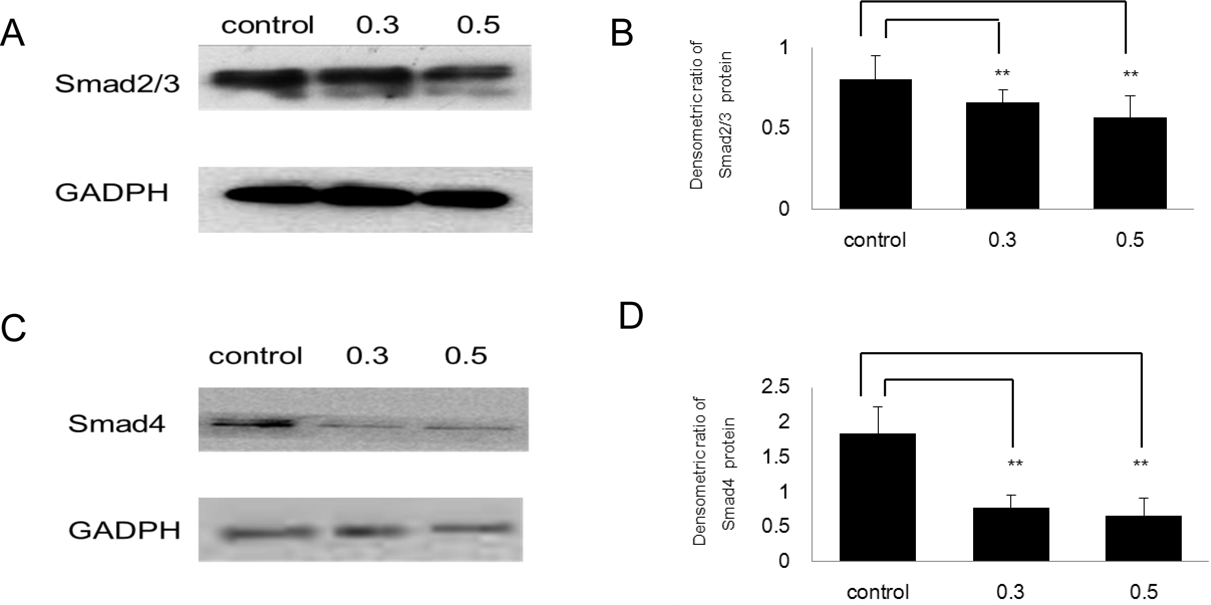
 Figure 5 of
Wang, Mol Vis 2013; 19:2626-2635.
Figure 5 of
Wang, Mol Vis 2013; 19:2626-2635.