Figure 4. Pirfenidone (PFD) inhibited expression of transforming growth factor beta 1 and transforming growth factor beta 2 in retinal
pigment epithelial cells. Retinal pigment epithelial (RPE) cells were treated in the absence or presence of PFD (control,
0.3 mg/ml or 0.5 mg/ml PFD) for 24 h. A and C: Reverse transcriptase (RT)–PCR and western blot analyses for transforming growth factor beta 1 (TGFβ1) were performed. E and G: RT–PCR and western blot results for detecting transforming growth factor beta 2 (TGFβ2) are shown. B, D, G, and H: The densitometry ratio data are presented. The densitometric ratio is the gene/protein of interest band density divided
by the glyceraldehyde-3-phosphate dehydrogenase (GADPH) band density. PFD inhibited the mRNA and protein levels of TGFβ1 and TGFβ2 in RPE cells (**n=3, the error bar indicates standard deviation [SD], p<0.01, relative to control).
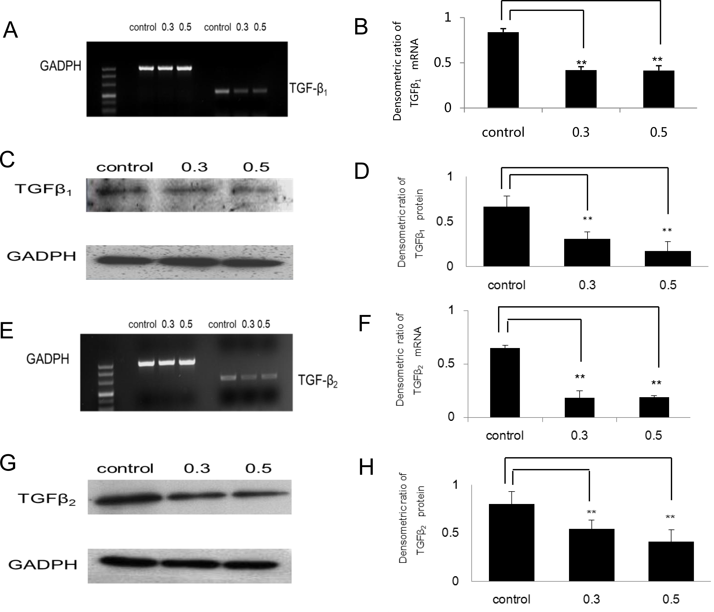
 Figure 4 of
Wang, Mol Vis 2013; 19:2626-2635.
Figure 4 of
Wang, Mol Vis 2013; 19:2626-2635.