Figure 3. Pirfenidone (PFD) inhibited expression of fibronectin in retinal pigment epithelial cells. Retinal pigment epithelial (RPE)
cells were treated in the complete medium (control) or 0.3 mg/ml or 0.5 mg/ml PFD for 24 h. Glyceraldehyde-3-phosphate dehydrogenase
(GADPH) was used to verify equal loading of genes and proteins. All experiments were repeated in triplicate, and the results
were reproducible. A and C: The expression of fibronectin (FN) were assayed using reverse transcriptase (RT)–PCR and western blot analyses. B and D: Compared with cells cultured in the complete medium, the densitometric ratios of FN mRNA and protein (normalized to GADPH)
in cells treated with PFD were significantly decreased (**n=3, the error bar indicates standard deviation [SD], p<0.01, relative
to control).
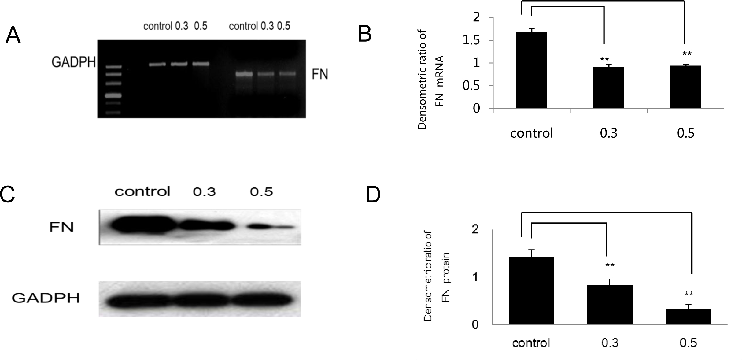
 Figure 3 of
Wang, Mol Vis 2013; 19:2626-2635.
Figure 3 of
Wang, Mol Vis 2013; 19:2626-2635.