Figure 2. Pirfenidone (PFD) inhibited expression of Snail1, α-smooth muscle actin, and connective tissue growth factor in retinal pigment
epithelial cells. Retinal pigment epithelial (RPE) cells were incubated for 24 h in complete medium (control), 0.3 mg/ml PFD,
or 0.5 mg/ml PFD. A: The expression of Snail1 was assayed with reverse transcriptase (RT)–PCR. C and E: Cells were harvested and subjected to western blot analysis with antibodies against connective tissue growth factor (CTGF)
and α-smooth muscle actin (α-SMA). The experiment was repeated at least three times. B, D, and F: The densitometry ratio data are presented. The densitometric ratio is the gene/protein of interest band density divided
by the glyceraldehyde-3-phosphate dehydrogenase (GADPH) band density. PFD inhibited expression of Snail1, α-SMA, and CTGF
in RPE cells. Samples significantly different from the control sample were indicated with symbols (**n=3, the error bar indicates
standard deviation [SD], p<0.01, relative to control).
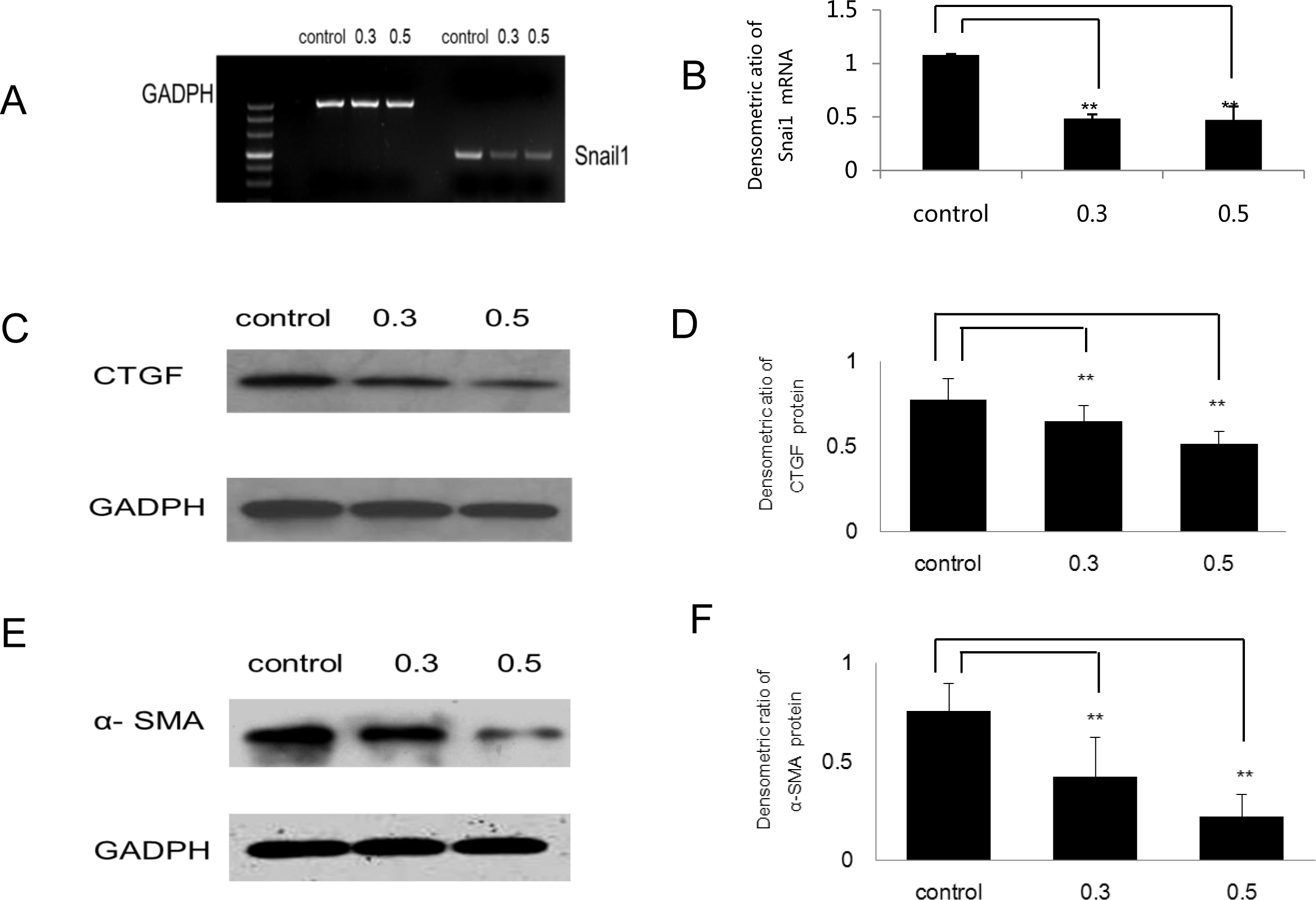
 Figure 2 of
Wang, Mol Vis 2013; 19:2626-2635.
Figure 2 of
Wang, Mol Vis 2013; 19:2626-2635.