Figure 2. Normal retinal structure is visible in tg21 nob mice. A: Retinal cross-sections obtained from 6-week old non-transgenic (left) and tg21 nob (right) littermates. The appearance of
the WT and tg21 nob retinas is comparable. B: Thickness of individual plexiform or cellular layers was assessed in WT and tg21 nob retinas. There was no significant difference
observable in any retinal layer. Bars indicate mean (± S.D. of 4 mice). Abbreviations: ONL represents the outer nuclear layer;
OPL represents the outer plexiform layer; INL represents the inner nuclear layer; IPL represents the inner plexiform layer;
GCL represents the ganglion cell layer.
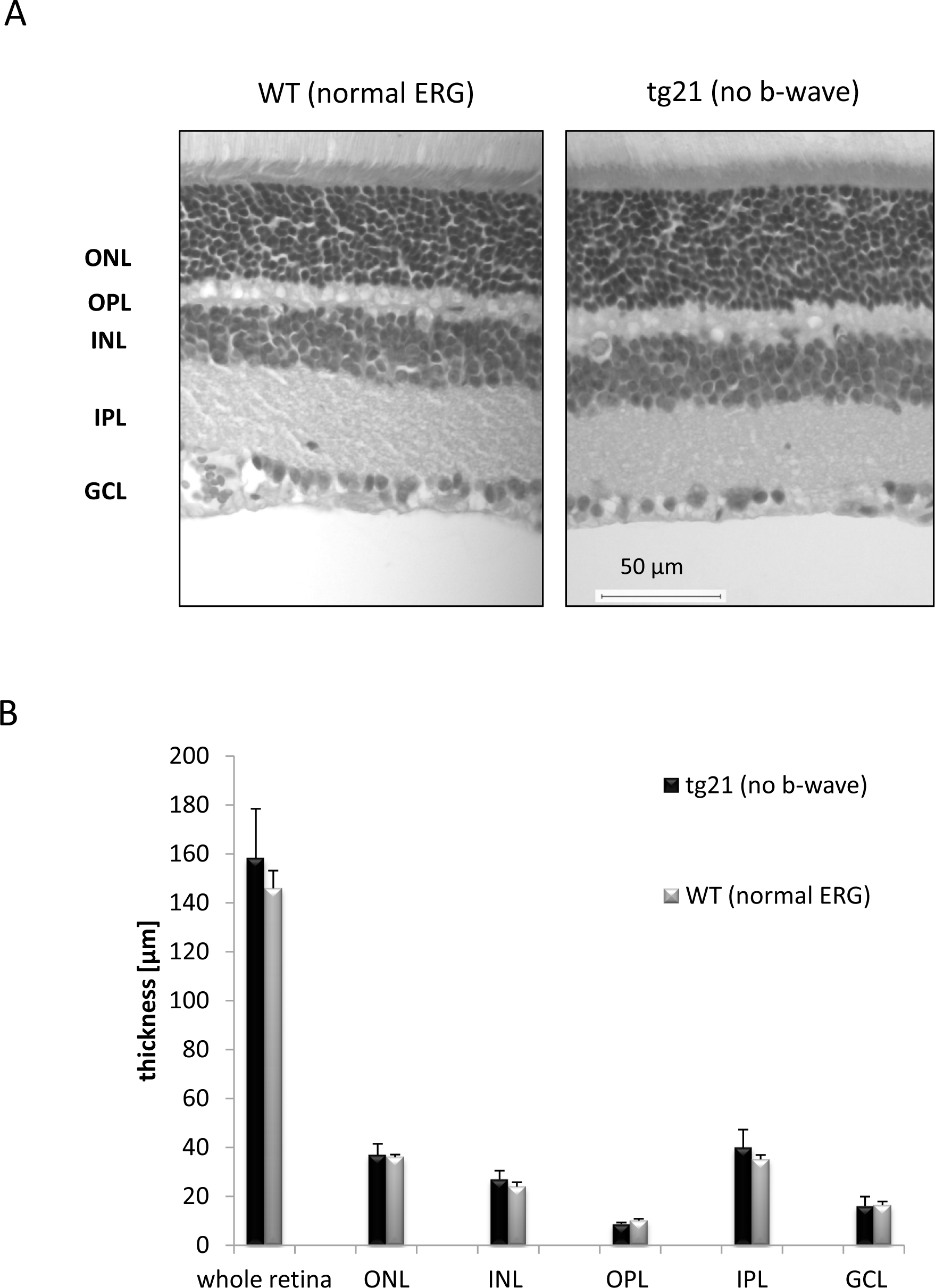
 Figure 2 of
Balmer, Mol Vis 2013; 19:2615-2625.
Figure 2 of
Balmer, Mol Vis 2013; 19:2615-2625.