Figure 4. Histopathological analysis of rat Fusarium keratitis. A–C illustrate histology of a corneal button from a representative control rat fitted with a contact lens soaked in saline. A, B: Hematoxylin and eosin (H&E) staining showed normal corneal caliber and absence of inflammatory infiltrates. C: Gomori methenamine silver (GMS) staining showed no fungal elements. D–F show a corneal button from a representative rat fitted with a contact lens soaked in Fusarium solani suspension with severe keratitis. D, E: H&E staining showed increased corneal thickness, severe inflammation, and necrotic stroma. F: A marked amount of fungal elements consistent with Fusarium were identified by GMS (red arrows). G–I illustrate a corneal button from a rat fitted with a contact lens soaked in Fusarium solution with mild to moderate keratitis. G, H: H&E staining showed increased corneal thickness, edema, and a moderate amount of polymorphonuclear neutrophils (yellow arrows).
I: A mild amount of fungal hyphae in the corneal stroma was identified with GMS (red arrows). D, G: Note the presence of endothelial plaques (black arrows). Scale bar=100 μm.
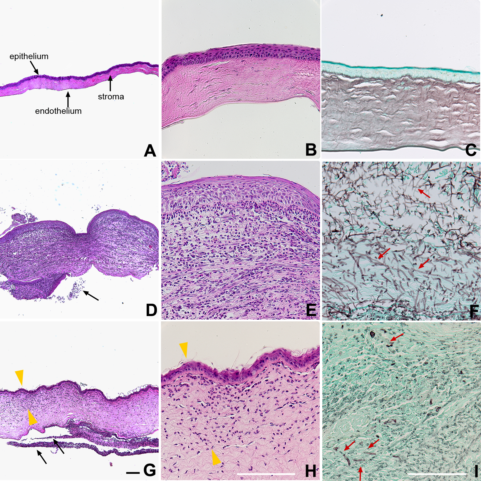
 Figure 4 of
Abou Shousha, Mol Vis 2013; 19:2596-2605.
Figure 4 of
Abou Shousha, Mol Vis 2013; 19:2596-2605.