Figure 1. The custom made contact lenses fitted perfectly on the rat eye. A: A representative slit-lamp picture illustrates the rat eye with the contact lens in place. The yellow arrowhead points to
the margin of the contact lens and the white arrow to the limbus. B: The corresponding optical coherence tomography image shows the rat eye with the contact lens in place. The yellow arrowhead
points to the contact lens and the white arrow to the cornea. Scale bar=100 μm.
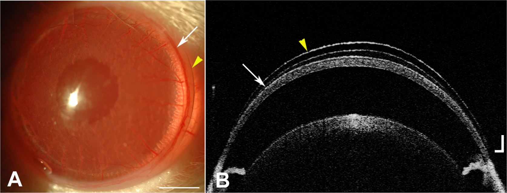
 Figure 1 of
Abou Shousha, Mol Vis 2013; 19:2596-2605.
Figure 1 of
Abou Shousha, Mol Vis 2013; 19:2596-2605.